1. A 28-year-old male patient complains of regurgitation, cough and heartburn that occurs every day after a meal, when bendi- ng forward or lying down. These problems have been observed for 4 years. Objective status and laboratory values are normal. FEGDS revealed endoesophagitis. What is the leading factor in the development of this disease?
2. A 70-year-old patient consulted a doctor about arrhythmic cardiac activity, dyspnea. Objectively: AP- 150/90 mm Hg, extrasystole arrhythmia (10-12 beats per minute), left ventricular systolic dysfuncti- on (ejection fraction at the rate of 42%). Which of antiarrhythmic drugs should be administered as initial therapy in this case?
3. A 57-year-old patient had an attack of retrosternal pain that lasted more than 1,5 hours. Objectively: the patient is inert, adynamic, has pale skin, cold extremities, poor volume pulse, heart rate - 120/min, AP- 70/40 mm Hg. ECG shows ST elevation in leads II, III, aVF. What condition are these changes typical for?
Explanation
This patient with ST segment elevation in leads II, III, aVF on ECG that also had an attack of retrosternal pain is definitely a case of STEMI - ST elevation Myocardial Infarction. With a blood pressure of 70/40mmHg (severe hypotension), the condition has deteriorated to a case of shock and in this case, it has a cardiac origin, therefore, it is a cardiogenic shock.
There is no arrhythmia recorded on ECG (no Atrial flutter or fibrillation, ventricular tachycardia or fibrillation), so it will be wrong to tag this an Arrhythmogenic shock. Perforated gastric ulcers can present with excessive bleeding and even result in shock but the patient will also present with hematemesis or melena and epigastric pain. Acute pericarditis can present with retrosternal pain but not likely to have shock. Acute pancreatitis will present with diarrhea, indigestion, epigastric belt-like pain radiating to the back, and it is unlikely for a patient with pancreatitis to have retrosternal pain and shock with the listed ECG findings.
4. A 63-year-old patient with persistent atrial fibrillation complains of moderate dyspnea. Objectively: peripheral edemata are absent, vesicular respiration is present, heart rate - 72/min, AP- 140/90 mm Hg. What combination of drugs will be most useful in the secondary prevention of heart failure?
Explanation
Beta blockers (drugs ending with the suffix ‘lol’ e.g. Metoprolol, propranolol, labetalol) can be used as antiarrhythmic drugs (treat arrhythmias like Atrial fibrillation). They belong to class II of the antiarrhythmic medications. They can also be used in hypertensive conditions. Beta 1 receptors are present in the heart and when a Beta blocker is deplored, it can reduce the heart rate thereby reducing the cardiac output and contractility. They can also reduce the transmission of impulses through the AV node which makes them useful as antiarrhythmic agents.
Angiotensin Converting Enzyme Inhibitors (ACEI) inhibit the conversion of Angiotensin I to Angiotensin II thereby inhibiting the vasoconstrictive properties of Angiotensin II. This makes them very useful as antihypertensives.
In this case, the question is interested in how to prevent heart failure and a combination of Beta blockers and ACE inhibitors are the most effective as they both reduce blood pressure and workload on the heart. They reduce both the preload and afterload of the heart which will prevent ventricular remodelling in the long run.
Cardiac glycosides tend to increase heart contractility and in the long run, this increase in workload can lead to hypertrophy. Diuretics can reduce the preload but not effectively reduce the afterload.
5. A 19-year-old patient complains of dyspnea on exertion. He often has bronchitis and pneumonia. Since childhood, the patient presents with cardiac murmur. Auscultation revealed splitting of the II sound above the pulmonary artery, systolic murmur in 3 intercostal space at the left sternal border. ECG showed right bundle branch block. What is the provisional diagnosis?
6. An emergency doctor has diagnosed a 32-year-old woman with generalized convulsive status epilepticus. The deterioration in the patient’s condition is caused by a sudden gap in the epilepsy treatment. Specify the doctor’s further tactics:
7. A 26-year-old patient with affective bipolar disorder has developed a condition manifested by mood improvement, behavioural and sexual hyperactivity, verbosity, active body language, reduced need for sleep. Which of the following drugs are most effective in this case?
Explanation
Neuroleptics, also known as antipsychotic medications, are used to treat and manage symptoms of many psychiatric disorders. They fall into two classes: first-generation or "typical" antipsychotics and second-generation or "atypical" antipsychotics." They are Dopamine D2 receptor antagonists. Antipsychotics are drugs that have a specific sedative effect, and which improve the attitude and calm the behavior of psychotic patients.
Typical antipsychotics: Haloperidol, Chlorpromazine
Atypical antipsychotics: Risperidone, Clozapine, Aripiprazole
Neuroleptic drugs are beneficial for the management of behavioral disorders. They are also associated with sedative effects, weight gain, or anticholinergic activity.
Bipolar disorder: a manic episode and depressed mood. For treatment of Bipolar disorders: Lithium, Valproic acid, Lamotrigine, Carbamazepine, and Atypical antipsychotics. This patient is currently having a manic episode and it's best to use a Neuroleptic (antipsychotic) with sedative effect to calm the patient down and to sleep.
A manic episode requires 3 or more of the following:
-
Decreased need for sleep
-
Distractibility
-
Impulsivity/Indiscretion: seeks pleasure without regard to consequences including sexual pleasure
-
Talkativeness or pressured speech
-
Grandiosity: inflated self esteem
-
Flight of ideas: racing thoughts
-
Increased goal oriented activity/psychomotor agitation
Antidepressants are used for Major Depressive Disorders; Tranquilizers are used for General Anxiety Disorder or Panic attacks.
8. A 48-year-old patient complains of weakness, subfebrile temperature, aching pain in the kidney region. These presentations turned up three months ago after hypothermia. Objectively: kidneys are painful on palpation, there is bi- laterally positive Pasternatsky’s symptom. Urine test res: acid reaction, pronounced leukocyturia, microhematuria, minor proteinuria - 0,165-0,33 g/l. After the urine sample had been inoculated on conventional media, bacteriuria were not found. What research is most required in this case?
Explanation
There are some characteristics in a urine examination that suggest a diagnosis of renal Tuberculosis, such as acid pH, leukocyturia and/or hematuria, associated with negative urine culture for the usual bacteria that causes urinary tract infection (bacteria were not found on conventional media).
Points worthy of note from the Urine test result: acid reaction, leukocyturia and after inoculation on conventional media, bacteriuria were not found.
Now, Mycobacterium tuberculosis is acid fast due to the presence of Mycolic acid, so they will produce an acid reaction. There is increased lymphocytes (leukocyturia) with M.tuberculosis and viral infection (Neutrophils will increase with bacteria - Neutrophilia). Also, M. tuberculosis will not grow on conventional media and they stain poorly with Gram staining because their cell wall has high lipid content. To detect M. tuberculosis, Ziehl-Neelsen stain (Carbol fuchsin) is used instead.
Nechiporenko: This method helps to determine the amount of cellular elements (WBC, RBC and casts) in 1 ml of urine.
Zimnitsky's test characterize the concentrating and excretory ability of the kidney.
Daily proteinuria monitoring will give information about the extent of nephrotic syndrome or any other nephropathy like hypertensive nephropathy.
Isotope renography is an imaging technique that’s used to check kidney function.
9. A 22-year-old vegetarian patient with signs of malnutrition consulted a doctor about smell and taste distortion, angular stomatitis. Objectively: expressively blue sclerae. The patient was diagnosed with iron deficiency anemia. What is the dominating clinical syndrome?
10. A week ago a 65-year-old patient suffered an acute myocardial infarction, his general condition deteriorated: he complains of dyspnea at rest, pronounced weakness. Objectively: edema of the lower extremities, ascites is present. Heart borders are extended, paradoxical pulse is 2 cm displaced from the apex beat to the left. What is the most likely diagnosis?
11. In a cold weather, the emergency room admitted a patient pulled out of the open water. There was no respiratory contact with the water. The patient is excited, pale, complains of pain, numbness of hands and feet, cold shiver. Breathing rate is 22/min, AP- 120/90 mm Hg, Ps- 110/min, rectal temperature is 34, 5oC. What kind of warming is indicated for this patient?
Explanation
Hypothermia is a severe condition in which the body temperature drops to an abnormally low level. It occurs when the body is unable to produce enough heat to counter the heat that it is losing. Under healthy conditions, the body maintains a relatively stable temperature of around 98.6˚F or 37˚C.
The following techniques can help treat hypothermia:
Passive external rewarming: This uses the individual’s heat-generating ability. It involves removing their cold, wet clothing, ideally replacing it with adequately insulated, dry clothing, and moving them to a warm environment.
Active external rewarming: This involves applying warming devices, such as hot-water bottles or warmed forced air, externally to truncal areas of the body. For example, the individual could hold a hot-water bottle under each arm.
Active core rewarming: This uses warmed, intravenous fluids to irrigate body cavities, including the thorax, peritoneum, stomach, and bladder. Other options include getting the individual to inhale warm, humidified air, or applying extracorporeal rewarming by using a heart-lung machine.
Do not give a person alcohol if they have signs of hypothermia, and avoid giving any drinks to an unconscious person.
From the vitals given, this patient is fairly stable except for the tachycardia (110bpm) and low body temperature, therefore passive warming will suffice. The patient was pulled out of the open water in a cold weather so removing the wet clothing, and replacing it with adequately insulated, dry clothing, and moving them to a warm environment will keep the patient warm.
12. Six months ago, a 5-year-old child was operated for CHD. For the last 3 weeks he has complained of fever, heart pain, aching muscles and bones. Examination results: \"white-coffee\" skin colour, auscultation revealed systolic murmur in the region of heart along with a noise in the III-IV intercostal space. Examination of fingertips revealed Janeway lesions. What is your provisional diagnosis?
13. A 58-year-old patient complains of a headache in the occipital region, nausea, choking, opplotentes. The presentations appeared after a physical exertion. Objectively: the patient is excited. Face is hyperemic. Skin is pale. Heart sounds are regular, the 2nd aortic sound is accentuated. AP- 240/120 mm Hg, HR- 92/min. Auscultation reveals some fine moist rales in the lower parts of the lungs. Liver is not enlarged. ECG shows signs of hypertrophy and left ventricular overload. What is the most likely diagnosis?
14. A 58-year-old patient complains of general weakness, loss of 10 kg of weight within 1,5 months, progressive pain in the lumbar region, increased blood pressure up to 220/160 mm Hg, subfebrile temperature. Objectively: in the right hypochondrium palpation reveals a formation with uneven surface and low mobility; veins of the spermatic cord and scrotum are dilated. Blood test results: Hb- 86 g/l, ESR- 44 mm/h. Urine test results: specific gravity - 1020, protein - 0,99 g/l, RBCs - cover the whole field of vision, WBCs - 4-6 in the field of vision. What is the provisional diagnosis?
15. After a contact with chemicals a plant worker has suddenly developed stridor, voice hoarseness, barking cough, progressing dyspnea. Objective examination reveals acrocyanosis. What is your provisional diagnosis?
16. A 40-year-old female patient complains of having a bulge on the anterior surface of neck for 5 years. Objectively: Ps- 72 bpm, arterial pressure - 110/70 mm Hg, in the right lobe of thyroid gland palpation reveals a mobile 4x2 cm node, the left lobe is not palpable, the basal metabolic rate is 6%. What is the most likely diagnosis?
17. A 40-year-old female patient has been hospitalized for attacks of asphyxia, cough with phlegm. She has a 4-year history of the disease. The first attack of asphyxia occurred during her stay in the countryside. Further attacks occurred while cleaning the room. After 3 days of inpatient treatment the patient’s condition has significantly improved. What is the most likely etiological factor?
18. 5 weeks after hypothermia a 22-year-old patient developed fever, weakness, muscle pain, inability to move independently. Objectively: tenderness, induration of shoulder and shin muscles, restricted acti- ve movements, erythema on the anteri- or surface of chest. There is a periorbi- tal edema with heliotropic erythema. Gottron’s sign is present. What study is required to verify the diagnosis?
19. A 24-year-old patient consulted a doctor about enlarged submandibular lymph nodes. Objectively: submandi- bular, axillary and inguinal lymph nodes are enlarged. Chest radiograph shows enlarged mediastinal lymph nodes. In blood: RBCs - 3,4 · 1012/l, Hb- 100 g/l, colour index - 0,88, thrombocytes - 190 · 109/l, WBCs - 7,5 · 109/l, eosinophils - 8%, stab neutrophiles - 2%, segmented neutrophiles - 67%, lymphocytes - 23%, ESR - 22 mm/h. What study is required to verify the cause of lymphadenopathy?
20. A patient operated for acute paraprocti- tis undergoes antibacterial and detoxi- fication therapy, the local course of the disease has the positive dynamics. Since the operation the patient has had chi- lls, pyrexia, tachycardia, euphoria for five days. The doctor suspected sepsis. What study will confirm the diagnosis?
21. A painter working at a motorcar plant has been diagnosed with moderately severe intoxication with ami- de compounds of benzene. The in-patient treatment resulted in a considerable health improvement. What expert deci- sion should be made in this case?
22. An 18-year-old patient since childhood suffers from bleeding disorder after minor injuries. His younger brother also has bleeding disorders with occasional haemarthrosis. Which laboratory test will be informative for diagnosis verification?
Explanation
Clotting time is the time required for a sample of blood to coagulate in vitro under standard conditions. The time taken for blood to clot mainly reflects the time required for the generation of thrombin. If the plasma concentration of prothrombin or of some of the other factors is low (or if the factor is absent, or functionally inactive), clotting time will be prolonged. The expected range for clotting time is 4-10 mins.
Clotting time involves the measurement of prothrombin time (PT) and activated partial thromboplastin time (aPTT). Prothrombin time assesses the extrinsic (factor VII) and common (factors X, V, II [prothrombin] and I [fibrinogen]) pathways, whereas aPTT assesses the intrinsic (prekallikrein, HMWK, and factors XII, XI, IX, and VIII) and common pathways.
23. A patient complains of fatigue, lack of appetite, pain and burning sensation in the tongue, numbness of the distal limbs, diarrhea. Objectively: pale skin with lemon-yellow tint, face puffiness, brown pigmentation in the form of a \"butterfly\", bright red areas on the tongue. The liver is 3 cm below the costal margin, soft. Blood count: RBCs - 1, 5 · 1012 /l, colour index - 1,2, WBCs - 3, 8 · 109 /l, thrombocytes - 180 · 109/l, eosinophils - 0%, stab neutrophils - 1%, segmented neutrophils - 58%, lymphocytes - 38% monocytes - 3%, RBC macrocytosis. ESR - 28 mm/h. What diagnosis are these presentation typical for?
24. Medical examination of a 19- year-old worker revealed generalized lymphadenopathy mainly affecting the posterior cervical, axillary and ulnar lymph nodes. There are multiple injection marks on the elbow bend skin. The man denies taking drugs, the presence of injection marks ascribes to influenza treatment. Blood count: RBCs- 3, 2·1012/l, Hb- 100 g/l, WBCs- 3, 1 · 109/l, moderate lymphopenia. What study is required in the first place?
25. During the periodic medical examination an assembly fitter (works on soldering details) didn’t report any health problems. Closer examination revealed signs of asthenic-vegetative syndrome. Blood included red blood cells with basophilic aggregations and a somewhat higher number of reticulocytes, urine had a high concentration of delta- aminolevulinic acid. The complex of symptoms indicates the initial stage of chronic intoxication with:
26. A 50-year-old patient complains of bursting pain in the left lower limb that is getting worse on exertion, swelling in the region of shin and foot. Objectively: left shin and foot are doughy, skin of the lower shin is indurated and has a bronze tint, subcutaneous veins are dilated, there is an ulcer with necrotic masses. What is the most likely diagnosis?
27. A 48-year-old patient was found to have diffuse enlargement of the thyroid gland, exophthalmia, weight loss of 4 kg in 2 months, sweating. Objectively: HR- 105/min, AP- 140/70 mm Hg. Defecati- on act is normal. What kind of therapy is recommended in this case?
28. A 48-year-old male patient complains of constant pain in the upper abdomen, mostly on the left, that is getting worse after taking meals; diarrhea, weight loss. The patient is an alcohol abuser. 2 years ago he had acute pancreatitis. Blood amylase is 4 g/h·l. Coprogram shows steatorrhea, creatorrhea. Blood glucose is 6,0 mmol/l. What treatment is indicated for this patient?
Explanation
This patient has a case of acute Pancreatitis. Recall that the pancreas has both endocrine and exocrine functions; for the exocrine part, it produces enzymes that aid in digestion such as amylase, lipase, trypsin. Absence of these enzymes will lead to symptoms such as steatorrhea (fats in feces), creatorrhea (undigested muscles in feces) etc. Panzinorm Forte is a combination of digestive enzymes. These enzymes are normally produced by the pancreas and are important for digesting fats, proteins, and sugars.
Panzinorm Forte is used to replace digestive enzymes when the body does not have enough of its own. Certain medical conditions can cause this lack of enzymes, such as cystic fibrosis, pancreatitis, pancreatic cancer, or pancreas surgery.
Panzinorm Forte may also be used to treat a condition called steatorrhea (loose, fatty stools).
Pirenzepine is an M1 selective antagonist used in treatment of peptic ulcer ( it reduces gastric acid secretion and muscle spasm). Contrykal is a protease inhibitor and is used in reducing blood loss in surgical procedures. Drotaverine is an antispasmodic drug used for cervical dilation in childbirth
29. A 20-year-old patient complains of severe headache, double vision, weakness, fever, irritability. Objectively: body temperature is at the rate of 38, 1oC, the patient is reluctant to contact, sensitive to stimuli. There is ptosis of the left eyelid, exotropia, anisocoria S>D, pronounced meningeal syndrome. On lumbar puncture the cerebrospinal fluid flowed out under a pressure of 300 mm Hg, the fluid is clear, slightly opalescent. 24 hours later there appeared the fibrinous film. Protein - 1,4 g/l, lymphocytes - 600/3 per mm3, sugar - 0,3 mmol/l. What is the provisional diagnosis?
30. A 32-year-old patient has developed an acute condition after hypothermia: temperature - 40oC, cough with 200 ml of sputum per day. The sputum is purulent, foul-smelling. To the right of the lower lobe the mixed moist rales can be auscultated. Blood tst results: WBCs - 18, 0 · 109/l, ESR - 45 mm/h. Radiographi- cally: in the lower lobe of the right lung there is a thick-walled cavity up to 6 cm in diameter with a high horizontal level. What is the most likely diagnosis?
31. A family consists of 5 persons. The husband is a stope miner. His spouse is a housewife. Their 20-year-old daughter works as a kindergarten teacher. Their 18-year-old son is a student. The grandmother is a pensioner, she has diabetes. Which member of this family can be primarily classed among the group of persons with a high risk of tuberculosis in the planning of preventive examinations for tuberculosis?
Explanation
Generally, persons at high risk for developing TB disease fall into two categories:
Persons in the second group include individuals with the following diseases; HIV infection (the virus that causes AIDS), Substance abuse, Silicosis, Diabetes mellitus, Severe kidney disease, Low body weight, Organ transplants, Head and neck cancer, Medical treatments such as corticosteroids or organ transplant, Specialized treatment for rheumatoid arthritis or Crohn’s disease.
32. A patient with suspected pheochromocytoma has normal blood pressure in the periods between the attacks and a tendency to tachycardia. Urine test revealed no pathology. It was decided to use a provocative test with histamine. What medication should be prepared to provide emergency care in case of a positive test result?
33. A 54-year-old patient complains of frequent painful urination, chills, fever up to 38*C. Urine test results: protein - 0,33 g/L, WBCs - up to 50-60 in the field of vision, RBCs - 5-8 in the field of vision, gram-negative bacilli. Which of the listed antibiotics should be preferred in this case?
34. Several hours before, a 28-year- old patient suddenly developed acute headache and repeated vomiting, then lost consciousness. Objectively: focal neurological symptoms were not found. Pronounced meningeal symptoms were revealed. AP - 120/80 mm Hg. According to clinical and liquorological findings the patient was diagnosed with subarachnoid haemorrhage. After administration of dehydrants the patient’s condition somewhat improved. What is the main component of further emergency care?
35. Gastric juice analysis of a 42-year- old male patient revealed absence of free hydrochloric acid at all stages. Endoscopy revealed pallor, thinning of gastric mucosa, smoothed folds. Microscopically the atrophy of glands with intestinal metaplasia was found. What disease is this situation typical for?
Explanation
The following types of chronic gastritis include;
Type A is caused by your immune system destroying stomach cells. And it can increase your risk of vitamin deficiencies, anemia, and cancer.
Type B, the most common type, is caused by Helicobacter pylori bacteria, and can cause stomach ulcers, intestinal ulcers, and cancer.
Type C is caused by chemical irritants like nonsteroidal anti-inflammatory drugs (NSAIDs), alcohol, or bile. And it can also cause stomach lining erosion and bleeding.
Observe that on examination, free HCL is absent and results from endoscopy and microscopy indicate the gastric cells have been destroyed - this occurs in Type A gastritis.
In menetrier's disease, we see an overgrowth of gastric cells ( foveola) found in the gastric lining which leads to the presence of large gastric folds.
36. A 24-year-old female teacher complains of dizziness and heart pain irradiating to the left nipple. Pain is not associated with physical activity and cannot be relieved by nitroglycerin, it abates after taking Valocordin and lasts an hour or more. The patient has a nearly 2-year history of this disease. Objectively: Ps- 76 bpm. AP- 110/70 mm Hg. Heart borders are normal, heart sounds are clear. The ECG shows respiratory arrhythmia. Radiograph of the cervicothoracic spine shows no pathology. Lungs, abdomen are unremarkable. What changes in blood formula can be expected?
37. A 60-year-old male patient, who works as a construction worker, complains of pain in the right hip and knee joints, that is getting worse on exertion. These presentations have been observed for the last 5 years. Objectively: the patient is overnourished. Right knee joint is moderately deformed. Examination of other organs and systems revealed no pathology. Blood test results: WBCs - 8, 2 · 109/l, ESR - 15 mm/h. Uric acid - 0,35 mmol/l. What is the most likely diagnosis?
38. A 32-year-old female complains of dizziness, headache, palpitation, tremor. For the last several months she has been under outpatient observation for the increased arterial pressure. Since recently such attacks have become more frequent and severe. Objectively: skin is covered with clammy sweat, tremor of the extremities is present. HR- 110/min, AP- 220/140 mm Hg. Heart sounds are muffled. Blood test results: WBCs - 9, 8 · 109/l, ESR - 22 mm/h. Blood glucose - 9,8 millimole/l. What disease is the most likely cause of this crisis?
Explanation
The woman not only shows signs of hypertension but also presents with changes relating to body metabolic activities (tremors, tachycardia, dizziness, palpitations, sweating - these are signs relating to stress ( caused by the release of stress hormones i.e. catecholamines). Pheochromocytoma is a tumor of the adrenal gland, it is characterised by the increased production of catecholamines ( adrenaline, noradrenaline) coupled with stress symptoms. Essential hypertension is also called primary hypertension- a form of hypertension with no underlying disease as its cause. Preeclampsia is a pregnancy-associated hypertension coupled with other signs. Primary hyperaldosteronism or Conn’s disease is also associated with the above stated signs , its other signs may include kidney related problems, excessive urination, low potassium content, flank pain etc.
39. A 55-year-old male has a 1,5-year history of viral cirrhosis with symptoms of portal hypertension. Over the last month the weakness has progrssed, there appeared coffee ground vomit. Fibrogastroduodenoscopy revealed variceal esophageal haemorrhage. What drug should be used to reduce the pressure in the portal vein?
40. A 40-year-old patient complains of fever up to 39*C, cough with sputum and blood admixtures, dyspnea, weakness, herpetic rash on the lips. Objectively: respiration rate - 32/min. Under the shoulder blade on the right the increased vocal fremitus and dullness of percussion sound were revealed. Auscultation revealed bronchial respiration. Blood count: WBCs - 14 · 109/l, ESR - 35 mm/h. What is the provisional diagnosis?
41. In the morning a patient had nausea, abdominal discomfort, single vomiting, dry mouth. In the evening, the patient presented with the increasing general weakness, double vision, difficult swallowing of solid food. Objectively: ptosis, mydriasis, anisocoria, absence of gag and pharyngeal reflex, dry mucous membranes. The previous evening the patient had dinner with canned food and alcohol. What is the presumptive diagnosis?
42. A 28-year-old female patient with a six-year history of Raynaud’s syndrome has recently developed pain in the small joints of hands, difficult movement of food down the esophagus. What kind of disease can you think of in this case?
Explanation
Systemic scleroderma is a chronic multisystem disorder of unknown etiology characterized clinically by thickening of the skin caused by accumulation of connective tissue and by involvement of visceral organs, including the gastrointestinal tract, lungs, heart, and kidneys. Common symptoms include Raynaud phenomenon, polyarthralgia, dysphagia, heartburn, and swelling and eventually skin tightening and contractures of the fingers.
In Periarteritis nodosa (PAN), small and medium sized vessels are inflamed leading to disruption in major organs such as nerves, intestinal tract, heart, and joints.
Systemic lupus erythematosus is characterised by a malar/ butterfly rash.
43. A 47-year-old patient came to see a doctor on the 7th day of disease. The disease developed very fast: after the chill body temperature rose up to 40*C and lasted up to 7 hours, then it dropped abruptly, which caused profuse sweat. There were three such attacks occuring once in two days. Two days ago the patient arrived from Africa. Objectively: pale skin, subicteric sclera, significantly enlarged liver and spleen. What is the cause of fever attacks in this disease?
44. On the 2nd day of illness a 27- year-old patient complains of unbearable headache, repeated vomiting. Objecti- vely: the patient is in a grave condition. He is conscious but adynamic. Lies in a forced position with his head thrown back. There is no skin rash. Nuchal muscles are evidently rigid, there are Kernig’s and Brudzinski’s signs. to- 39, 5oC, Ps- 120/min, AP- 130/80 mm Hg. The leading syndrome of this disease is caused by:
45. A 42-year-old female patient suffers from micronodular cryptogenic cirrhosis. Over the last week her condition has deteriorated: she developed convulsions, mental confusion, progressing jaundice. What study may give reasons for such aggravation?
46. A 60-year-old patient complains of recurrent pain in the proximal interphalangeal and wrist joints, their periodic swelling and reddening that have been observed for 4 years. X-ray picture represents changes in form of osteoporosis, joint space narrowing and single usuras. What is the most likely diagnosis?
47. A 57-year-old male patient complai- ns of dyspnea on exertion, heaviness in the right hypochondrium and shin edemata towards evening. Objectively: temperature - 38, 1oC, HR- 20/min, HR=Ps=92/min, AP- 140/90 mm Hg. There is apparent kyphoscoliosis. In the lungs single dry rales can be auscultated. Heart sounds are muffled, rhythmic. ECG: Rv1+Sv5=15 mm. X-ray picture shows the bulging of pulmonary artery cone, right ventricle enlargement. What is the most likely cause of this condition?
48. A 40-year-old woman with a history of combined mitral valve disease with predominant stenosis complains of dyspnea, asthma attacks at night, heart problems. At present, she is unable to do easy housework. What is the optimal tactics of the patient treatment?
49. A 25-year-old patient complains of having dull heart pain for the last 10 days, dyspnea on mild exertion, palpi- tations. The diasease developed 2 weeks ago after a respiratory infection. Objecti- vely: acrocyanosis, AP- 90/75 mm Hg, Ps- 96/min. Cardiac borders appear to be shi- fted to the left and right. Heart sounds are weak and have triple rhythm, there is systolic murmur at the apex. ECG showed sinus rhythm, complete left bundle branch block. What is the most likely diagnosis?
50. While staying in a stuffy room a 19- year-old emotionally labile girl developed severe weakness, dizziness, blackout, nausea and loss of consciousness without convulsions. Objectively: the patient is unconscious, the skin is pale, extremities are cold. AP- 90/60 mm Hg, Ps- 96/min, deficient, breathing is shallow. Pupillary and tendon reflexes are present. There are no pathological signs. What is the most likely diagnosis?
51. A patient complains of frequent, bulky, frothy stools with greenish mucus, crampi- ng pain in the umbilical region, abdomi- nal murmur, body temperature at the rate of 39o C . The patient associates the disease with consumption of soft-boiled eggs. What is the most likely pathogen?
52. X-ray picture of chest shows a density and an abrupt decrease in the upper lobe of the right lung. The middle and lower lobe of the right lung exhibit significant pneumatization. The right pulmonary hilum comes up to the dense lobe. In the upper and middle parts of the left pulmonary field there are multiple focal shadows. In the basal region of the left pulmonary field there are clear outlines of two annular shadows with quite thick and irregular walls. What disease is this X-ray pattern typical for?
53. A patient complains of impaired far vision. Previously his eyes often turned red and hurt. Objectively: the eyes are not irritated, the cornea is transparent, anterior chambers are median deep, their liquid is transparent. The iris of the right eye has not changed in colour, its pattern is unchanged. The pupil is of irregular shape, scalloped. Biomicroscopy of the crystalline lens reveals the areas of opacity and vacuoles. Make a diagnosis:
54. A 59-year-old male complains of heart pain, cough, fever up to 38*C . Three weeks ago he suffered a heart attack. Objectively: Ps- 86/min, rhythmic, blood pressure - 110/70 mm Hg. Auscultation reveals pericardial rub, rales beneath the shoulder blade. Radiography reveals no pathology. Blood count: WBCs - 10 · 109/l, ESR - 35 mm/h. ECG shows no dynamics. It would be most reasonable to administer the drugs of the following pharmaceutical group:
55. A 40-year-old female patient complains of headache, dizziness, muscle weakness, occasional cramps in the extremities. She has been taking anti-hypertensive medications for 10 years. AP- 180/100 mm Hg. Blood potassium - 1,8 millimole/l, sodium - 4,8 millimole/l. In urine: alkaline reaction, the relative density - 1012, protein and sugar are not found, WBCs - 3-4 in the field of vision, RBCs - 1- 2 in the field of vision. Conn’s syndrome is suspected. Which drug should be chosen for the treatment of arterial hypertension?
56. A 45-year-old patient, a sailor, was hospitalized on the 2nd day of the disease. A week ago he returned from India. Complains of body temperature of 41*C, severe headache, dyspnea, cough with frothy rusty sputum. Objectively: the patient is pale, mucous membranes are cyanotic, breathing rate is 24/min, tachycardia is present. In lungs: diminished breath sounds, moist rales over both lungs, crepitation. What is the most likely diagnosis?
57. A 53-year-old female patient complains of cardiac pain and rhythm intermissions. She has experienced these presentations since childhood. The patient’s father had a history of cardiac arrhythmias. Objectively: the patient is in grave condition, Ps- 220 bpm, AP- 80/60 mm Hg. ECG results: heart rate - 215/min, extension and deformation of QRS complex accompanied by atrioventricular dissociation; positive P wave. Some time later heart rate reduced down to 45/min, there was a complete dissociation of P wave and QRST complex. Which of the following will be the most effective treatment?
58. A 49-year-old patient complains of dyspnea, cough. There are no sputum discharges. He has repeatedly used salbutamol and intal but with no effect. Objectively: he is only able to sit while leaning on the table. Cyanosis of face, acrocyanosis are present. Breathing is shallow, laboured, in some parts it cannot be auscultated; there are diffuse rales, expiration is significantly prolonged. Heart sounds are muffled, tachycardia is present. Ps - 112/min, AP- 110/70 mm Hg. Liver is located near the costal arch. There are no peripheral edemata. What is your provisional diagnosis?
59. Blood typing resulted in positive isohemagglutination reaction with standard sera of А(II) and В(III) groups and negative reaction with sera of 0(I) and АВ(IV) groups. What is this result indicative of?
60. A patient complains of jerking, throbbing pain in the III finger on the right hand. The patient associates these pain onsets with an injury by a nail. The finger skin is hyperemic and tense, palpation with a bulbous-end probe reveals the most painful area. What is the provisional diagnosis?
61. A 24-year-old female patient complains of pain in the right hypochondrium that is getting worse after taking meals; nausea, fever up to 37,7*C, icteric skin, pain in the large joints. These presentations have been observed for 8 months. Objectively: hepatosplenomegaly. Blood test results: ESR- 47 mm/h, total bilirubin - 86,1 mmol/l, direct bilirubin - 42,3 mmol/l. Total protein - 62 g/l, albumins - 40%, globulins - 60%, gamma globulins - 38%. Viral hepatitis markers were not detected. The antibodies to smooth muscle cells are present. On ultrasound the portal vein diameter was of 1 cm. What is the most likely diagnosis?
62. A patient is 60 years old, retired, worked as deputy director of a research institute. Behavioural changes appeared 2 years ago after the death of her husband: she stopped looking after herself and leaving the house; then she refused to clean the apartment and cook. Mental status: temporal disorientation. The patient does not understand many of the questions, is confused; does not know how to cook soup or fasten a button. Her speech is characterized by stumbling and logoclonia. She does not recognize doctors, fellow patients. She cries a lot but can not explain the reason for tears. What is the mechanism of this pathology?
Explanation
Recall that the cerebral cortex is made up of different lobes, sulci and gyri that act as centres for various functions for example the occipital lobe for vision. The cerebral cortex can further be divided into brodmann's area with each area having a specific function eg area 44 & 45 (broca’s area) - for motor activities in speech production etc. This patient presents with temporal disorientation, problems with understanding questions, repetition of syllables (logoclonia), long term memory defects- these signs are mostly due to a defect in the Temporal lobe ( responsible for long term memory, processing auditory and visual sensory input, language recognition and formation of new memories). She also has defects with the cognitive function - can't clean or cook, fasten her button, confused etc.
Note that from anamnesis, since this patient does not have any underlying disease that can lead to an atherosclerotic formation, that option is ruled out. The above stated symptoms are indicative of a global cortex dysfunction affecting a wide area of the cerebral cortex and most likely due to the atrophy of the cerebral cortex.
63. A 27-year-old sexually active female complains of numerous vesicles on the right sex lip, itch and burning. Eruptions regularly turn up before menstruation and disappear 8-10 days later. What is the most likely diagnosis?
Explanation
Herpes simplex viruses are enveloped double stranded linear viruses. HSV-1 is also known as oral herpes ( gingivostomatitis, keratoconjunctivitis etc) while HSV-2 is known as genital or neonatal herpes. The clinical presentation of genital herpes include pain, itching, dysuria, vaginal and urethral discharge, tender lymphadenopathy, appearance of herpes vesicles on the external genitalia, labia majora, labia minora, vaginal vestibule - for women and glans penis, the prepuce, the shaft of the penis, and sometimes on the scrotum, thighs, and buttocks- for men.
Primary syphilis usually presents with a localized painless hard chancre on the genitals.
Cytomegalovirus is also known as human herpes virus-5, it is usually seen in immunocompromised patients and infected cells have characteristic ‘ owl's eye’ intranuclear inclusions.
64. A 26-year-old patient with left lower lobe pneumonia experiences an acute chest pain on the left during coughing. Objectively: diffuse cyanosis, extension of the left side of chest. Percussion reveals high tympanitis. Auscultation reveals no respiratory murmurs above the left side of chest. There is a deviation of the right cardiac border towards the midclavicular line. What examination will be the most informative?
65. After myocardial infarction, a 50- year-old patient had an attack of asthma. Objectively: bubbling breathing with frequency of 32/min, cough with a lot of pink frothy sputum, acrocyanosis, swelling of the neck veins. Ps- 108/min, AP- 150/100 mm Hg. Heart sounds are muffled. Mixed moist rales can be auscultated above the entire lung surface. What drug would be most effective in this situation?
66. Against the background of angina a patient has developed pain in tubular bones. Examination revealed generalized enlargement of lymph nodes, hepatolienal syndrome, sternalgia. In blood: RBCs - 3, 6 · 1012 /l, Hb- 87 g/l, thrombocytes - 45 · 109/l, WBCs - 13 · 109/l, blasts - 87%, stab neutrophils - 1%, segmented neutrophils - 7%, lymphocytes - 5%, ESR - 55 mm/h. What is the most likely diagnosis?
67. Examination of a 43-year-old man objectively revealed pallor of skin and mucous membranes, loss of tongue papillae, transverse striation of fingernails, cracks in the mouth corners, tachycardia. Blood test results: Hb- 90 g/l, anisocytosis, poikilocytosis. The most likely causative agent of this state is inadequate intake of:
Explanation
Iron deficiency anaemia is a condition where a lack of iron in the body leads to a reduction in the number of red blood cells. Iron is used to produce red blood cells, which help store and carry oxygen in the blood. If you have fewer red blood cells than is normal, your organs and tissues won't get as much oxygen as they usually would. Causes include blood loss ( bleeding, menorrhagia etc), poor diet, malabsorption and hookworms. Key findings include; koilonychia (spoon shaped, brittle nails), atrophic glossitis (loss of filiform papillae on the tongue), angular cheilitis (inflammation of mouth corners) etc. Note that Anisocytosis refers to the presence of RBCs of various sizes while poikilocytosis refers to RBCs of various shapes - both findings are usually in Iron deficiency anemias.
Copper deficiency will present with signs such as difficulty in learning and forming memories, weak and brittle bones, difficulty walking, vision loss (Kayser–Fleischer ring in eyes).
68. A patient had 4 generalized convulsive seizures within a day. Between the seizures the patient did not maintain clear consciousness (was in a coma or stupor). Specify his state:
Explanation
Status epilepticus (SE) is a common, life-threatening neurologic disorder that is essentially an acute, prolonged epileptic crisis. Status epilepticus is defined as a continuous seizure lasting more than 30 min, or two or more seizures without full recovery of consciousness between any of them. Based on recent understanding of pathophysiology, it is now considered that any seizure that lasts more than 5 min probably needs to be treated as SE.
It can also be defined as a seizure that persists for a sufficient length of time or is repeated frequently enough that recovery between attacks does not occur. Therefore, the key phrase here is “recovery between attacks does not occur.” The question puts it this way - ‘between the seizures the patient did not come to waking consciousness.’
Prolonged Status epilepticus can lead to cardiac dysrhythmia, metabolic derangements, autonomic dysfunction, neurogenic pulmonary edema, hyperthermia, rhabdomyolysis, and pulmonary aspiration. Permanent neurologic damage can occur with prolonged SE.
69. A 45-year-old patient complains of fever up to 40o C , general weakness, headache and spasmodic contraction of muscles in the region of a shinwound. The patient got injured five days ago when ti- lling soil and didn’t seek medical attenti- on. What kind of wound infection can be suspected?
70. A 60 year-old female has been suffering weakness, dizziness, fatigue over the last year. Recently she has also developed dyspnea, paresthesia. Objectively: skin and mucous membranes are pale and slightly icteric. The tongue is smooth due to the loss of lingual papillae. Liver and spleen are located at the costal margin. Blood count: Hb- 70 g/l, RBCs - 1, 7·1012/l, colour index - 1,2, macrocytes. Administer the patient a pathogenetically justified drug:
71. A 57-year-old female complains of having a sensation of esophageal compresion, palpitation, difficult breathing during eating solid food, occasional vomiting with a full mouth, \"wet pillow\" sign at night for the last 6 months. Objectively: body temperature - 39*C, height - 168 cm, weight - 72 kg, Ps- 76/min, АP- 120/80 mm Hg. X-ray revealed a considerable dilation of esophagus and its constriction in the cardial part. What pathology is most likely to have caused dysphagia in this patient?
72. A 49-year-old patient consulted a doctor about difficult swallowing, voice hoarseness, weight loss. These symptoms have been gradually progressing for the last 3 months. Objectively: the patient is exhausted, there are enlarged supraclavicular lymph nodes. Esophagoscopy revealed no oesophageal pathology. Which of the following studies is most appropriate in this case?
ExplanationA CT scan of the chest can help find problems such as infection, lung cancer, blocked blood flow in the lung (pulmonary embolism), and other lung problems. It also can be used to see if cancer has spread into the chest from another area of the body. Note that this patient most likely has cancer since there has been loss of weight over 3 months, progressing symptoms coupled with enlarged lymph nodes around this area. A lung x-ray gives a pictorial view of the heart, lungs, airways, blood vessels and the bones of the spine and chest but a CT scan is more detailed. Because it is able to detect very small nodules in the lung, chest CT is especially effective for diagnosing lung cancer at its earliest, most curable stage. Imaging of the esophagus is not needed as the question clearly stated that the esophagus revealed no pathology.
73. A 45-year-old female patient complaining of general weakness, nausea and vomiting has been delivered to a hospital by the ambulance. Recently there has been a lack of appetite, weight loss. Objectively: hyperpigmentation of skin, blood pressure at the rate of 70/45 mm Hg, bradycardia. Additional studies revealed the reduced concentration of aldosterone and cortisol in blood, decreased excretion of 17-ketosteroids and 17-oxyketosteroids in the urine, hyponatremia, chloropenia, hypokalemia. What therapeutic measures are required?
74. A 23-year-old female patient has a mental disease since the age of 18, the course of disease has no remission periods. At a hospital the patient mostly presents with non-purposeful foolish excitation: she makes stereotypic grimaces, exposed, masturbating in front of a loud laugh, repeating the stereotypical abusive shouts. The patient should be assigned:
75. A young woman with seborrhea adiposa has numerous non-itchy light brown and white spots with clear outlines and defurfuration on the torso and shoulder skin. What is the provisional diagnosis?
76. An emergency physician arrived to provide medical care for a hangman taken out of the loop by his relatives. The doctor revealed no pulse in the carotid arteries, absence of consciousness, spontaneous breathing and corneal reflexes; cadaver spots on the back and posterior parts of extremities. A person can be declared dead if the following sign is present:
Explanation
Forensic pathologists are essentially required to estimate the time since death (TSD) to assist in death investigation. The early post-mortem phase is most frequently estimated using the classical triad of post-mortem changes – rigor mortis, algor mortis and livor mortis.
Algor Mortis: Humans are warm-blooded organisms, which means that we maintain a constant internal temperature, regardless of the outside environment. The brain is our thermostat and the circulatory system is the main heat dissipator. However, within seconds of death, the brain cells begin to die and the heart stops pumping blood. Without the brain and and the blood distributing heat, the corpse eventually starts to match the outside temperature.
Rigor mortis is the post-mortem stiffening of muscles, caused by the depletion of adenosine triphosphate (ATP) from the muscles.
Livor Mortis: This is the final stage of death. When the heart stops beating, the blood is now at the mercy of gravity. It tends to collect at certain parts of the body. Depending on the position of the body, these parts would vary. For instance, if the person was flat on their back when they died, the blood would collect in the parts that are touching the ground (just as in this patient - cadaver spots were on the back and posterior parts of the extremities). If the person was hanging, it would collect in their fingertips, toes, and earlobes. Lividity starts with the skin where the blood has settled developing a bright red color. After a few hours, the color changes from red to bluish-purple. The bluish coloring of the skin is called livor mortis or cadaver spots.
Absence of spontaneous breathing, pulselessness or unconsciousness can be as a result of collapse (syncope) and not outright death. Absence of corneal reflexes is very possible and can be diagnosed in patients that are alive but with cranial nerve disorders (especially cranial nerve V and VII).
77. A 40-year-old patient is registered in a narcological dispensary. Somatically: skin is dramatically hyperemic, sclera are injected, hyperhidrosis is present. AP- 140/100 mm Hg, heart rate - 100/min. Mental state: autopsychic orientation is intact, allopsychic orientation is distorted. The patient presents with motor anxiety. There is a look of fear on his face. He refuses to talk about his problems and asks to release him immediately, because he \"may be killed.\" This state developed a day after a regular drinking bout. What is your provisional diagnosis?
78. A 35-year-old female patient has gained 20 kg weight within a year with the normal diet. She complains of chill, sleepiness, shortness of breath. The patient’s mother and sister are corpulent. Objectively: height - 160 cm, weight - 92 kg, BMI - 35,9. Obesity is uniform, there are no striae. The face is amimic. The skin is dry. The tongue is thickened. Heart sounds are muffled. HR- 56/min, AP- 140/100 mm Hg. The patient has constipations, amenorrhea for 5 months. TSH- 28 mkME/l (normal rate - 0,32-5). Craniogram shows no pathology. What is the etiology of obesity?
Explanation
Your thyroid produces thyroid hormone, which controls many activities in your body, including how fast you burn calories and how fast your heart beats. Diseases of the thyroid cause it to make either too much or too little of the hormone. Depending on how much or how little hormone your thyroid makes, you may often feel restless or tired, or you may lose or gain weight. Women are more likely than men to have thyroid diseases, especially right after pregnancy and after menopause.
Hypothyroidism is when your thyroid does not make enough thyroid hormones. It is also called underactive thyroid. This slows down many of your body's functions, like your metabolism. Signs and symptoms include the following: Feeling cold when other people do not; Constipation; weight gain, even though you are not eating more food; Feeling sad or depressed; Feeling very tired; Pale, dry skin; Dry, thinning hair; Slow heart rate (bradycardia); puffy face; hoarse voice; abnormal menstrual bleeding etc.
Note that the craniogram shows no pathology so we can rule out Hypothalamic-pituitary. Hypo-ovarian can present with amenorrhea, vaginal dryness, monophasic cycles etc but not with bradycardia or weight gain. Absence of abdominal striaes or skin hyperpigmentation and sometimes diabetes - rules out the option of cushing’s disease (hypercorticoidism).
The increased TSH levels seen in this patient implies an activation of the feedback mechanism signalling the body to produce more thyroid hormones; T3 and T4.
79. What juice should be included in a complex drug and dietary therapy for patients with gastric ulcer or duodenal ulcer and increased gastric juice acidity in order to accelerate the ulcer healing?
80. A 43-year-old alcohol abuser had not consumed alcohol for the last two days. In the evening he claimed to see rats and feel like they bite his feet. The patient is disoriented, agitated, all the time attempts to run somewhere. Specify the psychopathological syndrome:
81. A 67-year-old male patient complains of rash, severe pain in the subscapular region on the right. Objectively: skin in the right subscapular region is covered with linearly arranged pink-red edematous lesions that are somewhat infiltrated, and have clear boundaries. On the lesion surface there are vesicles with transparent exudate. What is the most likely diagnosis?
82. A 64-year-old male patient has a 35- year history of chronic pancreatitis. In the last 5 years, he claims to observe the pain abatement, bloating, frequent bowel movements up to 3-4 times a day, grayish, glossy stool with undigested food rests, the progressive loss of body weight. Change of symptoms in the patient is due to overlay of:
83. During the doctor’s round, a 56-year-old male patient with decompensated cirrhosis complains of dizziness, palpitations, moving black specks seen before the eyes, general weakness. The patient is pale, Ps- 110/min, AP- 90/50 mm Hg. What complication is most likely to have occurred in the patient?
84. A 57-year-old female patient complains of intense pain in the right hypochondrium irradiating to the right supraclavicular region. Skin and sclerae are icteric. There is tension and tenderness in the right hypochondrium on palpation. Body temperature is 38,8oC. Blood test results: WBC- 11, 2 · 109 /l, total bilirubin - 112 mmol/l (conjugated - 86 mmol/l, unconjugated - 26 mmol/l). What is the most likely diagnosis?
85. Examination of a 35-year-old patient with rheumatism revealed that the right heart border was 1 cm displaced outwards from the right parasternal line, the upper border was on the level with inferior margin of the 1st rib, the left border was 1 cm in from the left midclavicular line. Auscultation revealed atrial fibrillation, loud apical first sound, diastolic shock above the pulmonary artery. Echocardioscopy revealed abnormal pattern of the mitral valve motion. What heart disease is characterized by these symptoms?
86. A 54-year-old male patient works as an engineer. At the age of 35, he got infected with syphilis and treated it with \"traditional remedies\". About 5 years ago, he became forgetful, unable to cope with work, told cynical jokes, bought useless things, collected cigarette butts in the street. Objectively: the patient is indifferent, has slow speech, dysarthria, can make only primitive judgments, is unable to perform simple arithmetic operations or explain simple metaphors. The patient is untidy, takes no interest in anything, passive. Considers himself to be completely healthy. Qualify mental condition of the patient:
87. 5 days before, a 26-year-old female patient developed an acute condition. Objectively: marked headache, vomiting, weakness, poor appetite, temperature up to 39oC. Objectively: the patient is in a moderately grave condition, excited. The face is hyperemic, sclerae are injected. The tongue is coated with brown fur. The trunk and limbs are covered with plentiful roseolous and petechial rash. Hepatosplenomegaly is present. Complement binding reaction with Rickettsia prowazekii is positive with the titer of 1:640. What drug should be administered?
88. A 39-year-old female patient complains of dyspnea when walking, palpitation, edemata in the evening. The patient’s height is 164 cm, weight - 104 kg. Objectively: overnutrition. Heart sounds are weak, and tachycardia is present. The menstrual cycle is not broken. Blood sugar is 5,6 mmol/l, ACTH-response tests revealed no alterations. X-ray of the Turkish saddle revealed no pathology. What disease is it?
89. A 26-year-old male patient complains of a rash on the upper lip skin, which arose on a background of influenza with high-grade fever and is accompanied by pain and burning. The rash has been present for 3 days. Objectively: the skin of the upper lip is edematic and erythematous, grouped vesicles are filled with serous fluid and have a rough surface. What is the most likely diagnosis?
90. A 36-year-old female has a 7-year history of pollen allergy. Over the last 2 years in August and September (during ragweed flowering), the patient has had 2-3 asthma attacks that could be treated with one dose of salbutamol. Objectively: body temperature - 36, 5o C , respiratory rate - 18/min, Ps- 78/min, AP- 115/70 mm Hg. There is vesicular breathing above the lungs. Cardiac sounds are sonorous, of regular rhythm. What drug would be most effective to prevent asthma attacks during the critical season for the patient?
91. A 25-year-old female patient complains of marked weakness, sleepiness, blackouts, di- zziness, taste disorder. The patient has a hi- story of menorrhagia. Objectively: the pati- ent has marked weakness, pale skin, cracks in the corners of mouth, peeling nails, systolic apical murmur. Blood test results: RBC - 3, 4 · 1012 /l, Hb- 70 g/l, color index - 0,75, platelets - 140 · 109 /l, WBC- 6, 2 · 109 /l. What is the most likely diagnosis?
92. A 49-year-old male patient who had been scheduled for a surgery for gastric cancer underwent preoperative infusion therapy. Up to 3,0 liters of liquid was introduced into the right cubital vein. The following day, he felt a dragging pain in the right shoulder. Objectively: on the inner surface of the shoulder there is a longitudinal hyperemic zone, edema of skin, a tender cord. What complication occurred in the patient?
93. A 36-year-old female patient complains of intense pain in the knee joints and neck. In the morning she experiences pain in the interscapular region and leg joints; pain subsides after warm-up gymnastics. The patient is overnourished, there is a clicking sound in the knees when squatting, the knees are somewhat disfigured, painful on palpation. Blood test results: ESR- 18 mm/h, WBC- 8,0 · 109 /l. Radiography reveals subchondral sclerosis in the left knee. What is the basis of this pathology?
94. A 35-year-old female reports heart pain (aching and drilling) occurring mainly in the morning in autumn and spring and irradiating to the neck, back and abdomen; rapid heartbeat; low vitality. Occurrence of this condition is not associated with physical activity. In the evening, the patient’s condition improves. Study of somatic and neurological status, and ECG reveal no pathology. What pathology is most likely to have caused these clinical presentations?
95. A 33-year-old female complains of escalating spastic pain in the abdomen after the psycho-emotional stress. The patient has intermittent bowel movements, that is 2-3 bowel movements after waking up alternate with constipation lasting for 1-2 days. Objectively: body weight is unchanged, there is moderate pain on palpation of the sigmoid colon. Hb- 130 g/l, WBC- 5,2 · 109/l, ESR- 9 mm/h. Proctosigmoidoscopy causes pain due to spastic bowel condition, intestinal mucosa is not changed. In the lumen there is a lot of mucus. What is the most likely diagnosis?
96. After a holiday in the Crimea, a 36-year-old female patient presents with severe pain in the elbow joints, dyspnea and weakness. The body temperature is of 37,6*C, the skin is pale, there is erythema of cheeks and nose, lower lip ulceration. Visual inspection reveals no changes in the joints, the right elbow movement is limited. There is murmur and pleural friction in the lungs below the right angle of the scapula. Cardiac sounds are muffled, there is tachycardia, gallop rhythm, Ps- 114/min. AP- 100/60. What is the most likely diagnosis?
Explanation
The key finding here is the erythema on the cheeks and bridge of nose which describes a malar/ butterfly rash classically seen in patients with Systemic lupus erythematosus.
SLE is a chronic inflammatory disease of unknown cause which can affect the skin, joints, kidneys, lungs, nervous system, serous membranes and/or other organs of the body. Distinct immunologic abnormalities, especially the production of a number of antinuclear antibodies, other signs include, arthralgia, myalgia, nephritis, systolic murmurs in about 30% of patients, dry pleurisy in the lungs etc. The dry pleurisy is characterised by an abnormal sound on auscultation called a pleural friction rub.
Rheumatic carditis and arthritis will be observe in a case of rheumatic fever ( prior infection by group A beta hemolytic streptococci infection)
97. A 63-year-old male patient complains of cough with expectoration of mucous blood- streaked sputum, asthma, low-grade fever, general weakness. These presentations have been observed for 3 months. The patient has been a smoker since childhood. Objecti- vely: to- 37,4oC, respiratory rate is 26/min, Ps- 82/min, rhythmic. AP- 130/85 mm Hg. There is limited breathing movement in the right side of chest cavity, as well as percussive dullness and diminished breath sounds. Radi- ograph shows a homogeneous opacity of the pulmonary field on the right with the mediasti- num displacement to the affected side. What is the most likely diagnosis?
98. A 33-year-old male patient developed a condition that had a stormy clinical course: chills, fever up to 39oC, vomiting, epigastric pain, diarrhea with watery smelly feces. 6 hours before, he ate a raw egg, fried potatoes with stewed meat, drank some juice. What pathogen is likely to have caused this conditi- on?
99. A 76-year-old male consulted a therapist about slow discharge of urine with a small jet. The patient reported no cardiac problems. Examination revealed atrial fibrillation with a heart rate of 72/min and without pulse deficit. There are no signs of heart failure. ECG confirms the presence of atrial fibrillation. From history we know that the arrhythmia was detected three years ago. What tactics for the treatment of atrial fibrillation in the patient should be chosen?
100. A 53-year-old male has been admitted to a hospital for an attack of renal colic which has repeatedly occurred throughout the year. Objectively: in the region of auricles and the right elbow some nodules can be seen that are covered with thin shiny skin. Ps- 88/min, AP- 170/100 mm Hg. There is bilateral costovertebral angle tenderness (positive Pasternatsky’s symptom). The patient has been scheduled for examination. What laboratory value would be most helpful for making a diagnosis?
101. A 58-year-old female patient complains of spontaneous bruises, weakness, bleeding gums, dizziness. Objectively: the mucous membranes and skin are pale with numerous hemorrhages of various time of origin. Lymph nodes are not enlarged. Ps- 100/min, AP- 110/70 mm Hg. There are no changes of internal organs. Blood test results: RBC - 3,0·1012/l, Нb - 92 g/l, colour index - 0,9, anisocytosis, poikilocytosis, WBC - 10·109/l, eosinophils - 2%, stab neutrophils - 12%, segmented neutrophils - 68%, lymphocytes - 11%, monocytes - 7%, ESR - 12 mm/h. What laboratory test is to be determined next for making a diagnosis?
102. A 48-year-old male in-patient undergoes treatment for essential hypertension of II- B stage. It is known from history that he works in a design engineering office. His job involves neuro-emotional stress. Which of these foodstuffs do not stimulate the central nervous system and can be recommended for the patient?
103. A 48-year-old male patient complains of pain in the lower extremities, especially when walking, intermittent claudication, numbness in the fingers, cold extremities, inability to walk more than 100 meters. Sleeps with his leg lowered. The patient has been a smoker since he was 16, abuses alcohol, has a history of CHD. The left lower extremity is colder than the right one, the skin of extremities is dry, pedal pulse cannot be palpated, femoral pulse is preserved. What is the most likely diagnosis?
Explanation
Obliterating Endarteritis is also known as Buerger’s disease and vasculitis. The condition is thought to occur as a result of tobacco use and more common in males. The disease is characterized by inflammation and thrombosis of small and medium arteries of the legs and feet which recurs and progresses in stages. During the ischemic period, the veins of the lower leg are inflamed, skin has ulceration, and there is gangrene with intermittent claudication. During the nutritional disorder period there is persistent pain and muscle atrophy. During the necrosis period, the pain becomes worse as infection leads to the development of ulcers and gangrene. Pains usually persist; In addition, the patient usually complains of coldness and numbness. Burning sensations is another disturbing symptom.
Treatment usually has to do with smoking cessation.
Raynaud disease is a condition in which some areas of the body feel numb and cool in certain circumstances. In Raynaud's phenomenon, smaller arteries that supply blood to the skin constrict excessively in response to cold, limiting blood supply to the affected area. The fingers, toes, ears and tip of the nose are commonly involved and feel numb and cool in response to cold temperatures or stress.
Leriche Syndrome, also commonly referred to as aortoiliac occlusive disease, is a product of atherosclerosis affecting the distal abdominal aorta, iliac arteries, and femoral popliteal vessels. Its triad include; claudication, erectile dysfunction and decreased distal pulse.
104. A 38-year-old male complains of tonic tension of the masticatory muscles, so that he cannot open his mouth. 12 days before, he was bitten by an unknown dog. Objectively: there is pronounced tension and twitching of the masticatory muscles. What is the most likely diagnosis?
105. A 72-year-old male had had a moderate headache. Two days later, he developed the progressing speech disorders and weakness in the right extremities. The patient has a history of myocardial infarction, arrhythmia. Study of the neurologic status revealed elements of motor aphasia, central paresis of the VII and XII cranial nerves on the right, central hemiparesis on the same side and hyperaesthesia. What is the most likely diagnosis?
106. 2 weeks after having quinsy, a 26-year-old male patient got facial edemata, moderate pain in the sacrum. Objectively: body temperature is 37.5*C, AP- 100/80 mm Hg. Urinalysis results: RBC- up to 100 fresh cells in per HPF, protein - 2,2 g/l, hyaline cylinders - up to 10 per HPF, relative density - 1002. What is the most likely diagnosis?
107. A 27-year-old patient has a severe headache, nausea and vomiting. Objectively: body temperature is 38.9*C, there is a haemorrhagic stellate rash on the legs. The patient takes meningeal pose in bed. Meningeal symptoms are strongly positive. Deep reflexes are brisk, uniform. Pathological reflexes are absent. It has been suspected that the patient has epidemic cerebrospinal meningitis. Which of additional tests should be performed in the first place to verify the diagnosis?
108. 3 hours before, a 68-year-old male pati- ent got a searing chest pain radiating to the neck and left forearm, escalating dyspnea. Ni- troglycerin failed to relieve pain but somewhat reduced dyspnea. Objectively: there is cri- mson cyanosis of face. Respiratory rate is 28/min. The patient has vesicular breathing with isolated sibilant rales. Heart sounds are muffled, with a gallop rhythm. Ps- 100/min, AP- 100/65 mm Hg. ECG shows negative T- wave in V 2 − V 6 leads. What drug can reduce the heart’s need for oxygen without aggravati- ng the disease?
109. Two years ago, a 46-year-old patient was diagnosed with stage I silicosis. Currently the patient complains of escalating dyspnea, pain in the infrascapular regions. Radiograph shows a diffuse enhancement and distorti- on of lung markings, as well as multiple nodular shadows 2-4 mm in diameter. There is interlobar pleural density on the right. Dense shadows are found in the hilar regions. Specify the form of radiographic pulmonary fibrosis in this case:
110. A 63-year-old female complains of general weakness, a feeling of heaviness, compression in the epigastrium, postprandial fullness, nausea, belching after meals. These symptoms have been observed for about 15 years. Objectively: body temperature is 36,4*C , respiratory rate - 20/min, Ps - 88/min, blood pressure - 115/75 mm Hg. Skin and mucous membranes are pale. Blood test results: RBC - 2,0·1012/l,Hb-100g/l. Tests revealed parietal- cell antibodies. What is the most likely reason for the development of anemia in this patient?
111. During dressing of a poorly-granulating wound Pseudomonas aeruginosa infection was revealed. What medication would be optimal for the wound d-bridement?
112. A 30-year-old female patient has been delivered to a hospital for sudden dyspnea progessing to asthma, sensation of having a \"lump in the throat\", hand tremor, fear of death. The attack has developed for the first time and is associated with a strong emotion. There is no previous history. Objectvely: respiratory rate - 28/min, Ps- 104/min, rhythmic, AP- 150/85 mm Hg. The patient has rapid superficial vesicular breathing with extended expiration. Percussion findings: heart borders are not changed. Cardiac sounds are loud, rhythmic. What is the most likely diagnosis?
113. A 42-year-old male patient wth essential hypertension presents with headache, palpitations, unexplained fear. Objectively: Ps- 100/min, AP- 200/100 mm Hg, the left border of cardiac dullness is displaced by 1,5 cm to the left, vesicular breathing is present. ECG shows sinus tachycardia, signs of left ventricular hypertrophy. What drug should be administered as an emergency?
114. A 37-year-old male patient has a history of diabetes of moderate severity. On the left side of face the patient has a carbuncle. What severe complication might have occurred in the patient?
115. A 42-year-old female patient complains of a dull pain in her left side, low-grade fever, accelerated painful urination in small portions. These presentations have been observed for three years. For a long time, the patient has had cystitis with frequent exacerbations, there is pulmonary tuberculosis in the past history. Urinalysis results: microscopic hematuria, leukocyturia. What is the most likely provisional diagnosis?
116. A 65-year-old male patient complains of dyspnea that is getting worse with exertion, morning cough with expectoration of mucous sputum. For about 15 years, he has been subject to regular medical check-up for chronic bronchitis. The patient takes berodual (16 inhaled doses per day). Objectively: body temperature is 36, 8o C , RR- 24/min, Ps- 110/min, AP- 145/90 mm Hg. Auscultation reveals a lot of dry rales above the lungs. FEV1- 65%. What is the optimal tactics of further management of the patient?
117. A 49-year-old countryman got an itching papule on the dorsum of his right hand. In the centre there is a vesicle with serosanginous exudate. Within the next 2 days the patient developed a painless edema of hand and forearm. On the 4th day the temperature rose to 38,5*C, in the right axillary region a large painful lymph node was found. One day before the onset of the disease the patient had examined a dead calf. What is the most likely diagnosis?
Explanation
Bacillus anthracis is an aerobic, non-motile, spore forming, large non-hemolytic Gram-positive rod that grows well on blood agar. The clinical manifestations of human anthrax - Cutaneous and Inhalation.
Cutaneous anthrax, the most common form of naturally occurring disease, begins as a small, painless, pruritic papule that within 2 days enlarges, develops vesicles, and ulcerates to form an eschar. Inhalational anthrax follows inhalation of infectious doses of anthrax spores.
When anthrax spores get into the skin, usually through a cut or scrape, a person can develop cutaneous anthrax. Human cases may follow contact with contaminated carcasses (dead calf) or animal products. This can happen when a person handles infected animals or contaminated animal products like wool, hides, or hair. Cutaneous anthrax is most common on the head, neck, forearms, and hands. It affects the skin and tissue around the site of infection.
Bubonic plague is caused by bacteria called Yersinia pestis. It’s usually spread by fleas. These bugs pick up the germs when they bite infected animals like rats, mice, or squirrels.
118. A 38-year-old female suddenly developed acute inflammatory rash in form of roseolas, papules, vesicles that are scattered on the skin of trunk in irregular and predominantly focal manner. The rash appeared a few hours after visiting a restaurant. The patient complains of itching skin. What is the most likely diagnosis?
Explanation
Toxicodermatosis is a disease of the skin caused by a poison or by an allergen. The causative agents may be such chemical substances as antibiotics, sulfanilamides, and bromine, iodine, arsenic, and mercury compounds, or such food products as berries, cheese, or fish. As described in this scenario, the rash appeared after visiting a restaurant. Toxicoderma can also occur as a metabolic disorder or an occupational disease.
The causative agent enters the body through the respiratory organs or the digestive tract, during injections, or by direct contact with the skin. The primary manifestation of toxicoderma is a rash, which is generally symmetrical; it can take the form of inflamed or hemorrhagic maculae, papule, or vesicles (blisters). In severe cases, widespread areas of the skin are affected (erythroderma) and the patient manifests fever, cardiovascular disorders, or symptoms of anaphylactic shock.
With the removal of the allergen the symptoms of toxicoderma rapidly disappear, although relapses (usually severe) may occur with subsequent repeated exposure to the causative agent. Treatment consists in avoiding contact with the suspected allergen or toxic agent, as well as in the use of desensitizing agents and of expectant treatment.
Atopic dermatitis is a chronic condition that requires symptom management. It’s characterized by an itchy, red rash that usually appears at joints in your body, such as knees or elbows, and even around the neck. This condition occurs in flare-ups or bouts, meaning that it gets worse and improves in irregular cycles.
Many doctors use the terms “atopic dermatitis” and “eczema” interchangeably. They are both generic terms for “inflammation of the skin.” Eczema usually starts in early infancy, but also affects a substantial number of adults. It is commonly associated with elevated levels of immunoglobulin E (IgE) and a series of allergic diseases—including food allergy, asthma, and allergic rhinitis.
Contact Dermatitis occurs when your skin has a reaction to something it’s come in contact with. This can include bleach, soap, poison ivy, certain metals, or other irritants. The rash is typically red and may itch or burn.
119. After lifting a load, a 36-year-old male patient has experienced a severe pain in the lumbar region, which spread to the right leg and was getting worse when he moved his foot or coughed. Objectively: the long back muscles on the right are strained. Achilles jerk is reduced on the right. There is a pronounced tenderness of paravertebral points in the lumbar region. The straight leg raise (Lasegue’s sign) is positive on the right. What additional tests should be performed in the first place?
120. A 26-year-old female patient has an 11-year history of rheumatism. Four years ago she suffered 2 rheumatic attacks. Over the last 6 months there have been paroxysms of atrial fibrillation every 2-3 months. What option of antiarrhythmic therapy or tactics should be proposed?
121. On the second day of the disease a 22- year-old male patient complains of high-grade fever, headache in the region of forehead and superciliary arches, and during eye movement; aching muscles and joints. Objectively: body temperature is 39o C . Face is hyperemic, sclerae are injected. The mucous membrane of the soft palate and posterior pharyngeal wall is bright hyperemic and has petechial hemorrhages. What changes in the hemogram are typical for this disease?
122. A 44-year-old male patient complains of severe non-localized abdominal pain, pain in the right shoulder girdle, repeated vomiting, red urine. The onset of the disease is associated with alcohol consumption. The face is hyperemic. AP- 70/40 mm Hg. Abdominal radiography reveals no pathological shadows. Hemodiastase is 54 mg/h/l. Prothrombin is 46%. What is the provisional diagnosis?
123. A 41-year-old male patient was delivered to a hospital unconscious. During the previous 7 days he had been taking large doses of biseptolum for a cold. The night before, he began complaining of dyspnea, especially when lying down, swollen legs, 2-day urinary retention. In the morning he had seizures and lost consciousness. Objectively: noisy breathing at the rate of 30/min, edematous legs and lumbar region, Ps- 50/min. Plasma creatinine is 0,586mmol/l,plasmapotassium-7,2mmol/l. What treatment is necessary for this patient?
124. A 45-year-old male patient with acute abscess of the left lung has suddenly developed acute chest pain and dyspnea while coughing, tachycardia has increased. The control Ro-gram shows left lung collapse, the air in the left pleural cavity and a horizontal fluid level. What is the mechanism of this complication?
125. A 24-year-old male patient had been diagnosed with class III diffuse toxic goiter. There is moderate hyperthyroidism. A surgery was suggested, and the patient agreed to it. What preoperative measures should be taken for prevention of thyrotoxic crisis in the postoperative period?
Explanation
The patient in question already has diffuse toxic goiter with clinical manifestation of hyperthyroidism. Thyroid storm, also referred to as thyrotoxic crisis, is an acute, life-threatening, hypermetabolic state induced by excessive release of thyroid hormones in individuals with thyrotoxicosis. During a thyroid storm, an individual’s heart rate, blood pressure, and body temperature can soar to dangerously high levels. Without prompt, aggressive treatment, thyroid storm is often fatal. Thyroid storm may be precipitated by the stress of surgery, anesthesia, or thyroid manipulation and may be prevented by pretreatment with antithyroid drugs.
126. A 26-year-old male patient complains of pain in the right knee, which is getting worse in the morning. Two weeks before, he consulted an urologist about prostatitis. Objectively: conjunctivitis is present. There is also peri-articular edema of the knee joint, redness of the overlying skin. Rheumatoid factor was not detected. Until further diagnosis is specified, it would be reasonable to start treatment with the following antibiotic:
127. A 66-year-old female patient has been admitted to a hospital for massive gross hematuria with release of shapeless blood clots, frequent painful urination. The patient also reports a moderate weight loss within 3-4 months. Gross hematuria that was not accompanied by pain and dysuria first occurred three months ago for no apparent reason, and after a few days the bleeding subsided independently. What is the most likely diagnosis?
128. A 47-year-old female patient has an 8-year history of ulcerative colitis, has been treated with glucocorticoids. She complains of cramping pain in the umbilical region and left iliac region which has significantly increased during the past 2 weeks, diarrhea with mucus and blood 4-6 times a day, elevated body temperature up to 38 − 39*C, headache and pain in the knee joints. Objectively: the patient is in moderate condition, Ps- 108/min, AP- 90/60 mm Hg; heart and lungs are unremarkable; the tongue is moist; abdominal muscle tone is significantly decreased; peristaltic noises are absent. What complication developed in the patient?
Explanation
The term inflammatory bowel disease describes a group of disorders in which the intestines become inflamed. Two major types of inflammatory bowel disease are Ulcerative colitis and Crohn’s disease. Ulcerative colitis is limited to the colon or large intestine. Crohn’s disease, on the other hand, can involve any part of the gastrointestinal tract from the mouth to the anus.
The patient in question suffers from Ulcerative colitis. Common complications include; Toxic megacolon (toxic dilatation of the colon), or fulminant colitis. If Ulcerative colitis attack is severe, the colon might burst, or infection could spread through your body. Your intestines stop moving waste, and your belly swells. However, if a toxic megacolon is not urgently attended to, it could further lead to perforation.
Fistulas, abscess, and strictures are commonly associated with Crohn’s disease.
129. For 3 days, a 28-year-old female patient had had the body temperature increase up to 38*C, weakness, poor appetite, nausea, a single vomiting. On the 4th day the temperature was normal, the condition improved, but the jaundice developed. Objectively: moderate ictericity of skin, +3 cm enlarged liver of elastic consistency. Ortner’s, Kehr’s and Voznesensky’s symptoms are negative. What test will verify the diagnosis?
130. A 40-year-old male patient has had heaviness in the epigastric region for the last 6 months. He has not undergone any examinations. The night before, he abused vodka. In the morning there was vomiting, and 30 minutes after physical activity the patient experienced dizziness and profuse hematemesis. What pathology should be suspected in the first place?
131. A 55-year-old patient complains of severe itching, burning and pain in the eyes, skin redness in the outer corners of the palpebral fissure. Objectively: skin around the outer corners of the palpebral fissure is macerated, eczematous, there are single moist cracks. Palpebral conjunctiva is hyperemic, quaggy. There are minor discharges in form of stringing mucus. What is the most likely diagnosis?
Explanation
The conjunctiva is a thin, translucent, relatively elastic tissue layer with both bulbar and palpebral portions. The bulbar portion of the conjunctiva lines the outer aspect of the globe, while the palpebral portion covers the inside of the eyelids.
Conjunctivitis refers to any inflammatory condition of the membrane that lines the eyelids and covers the exposed surface of the sclera. It is the most common cause of “red eye.”
Chronic conjunctivitis is most commonly caused by Staphylococcus species, although other bacteria are occasionally involved. This type of conjunctivitis often develops in association with blepharitis - bacterial colonization of the eyelid margins (palpebral conjunctiva is hyperemic and quaggy). The symptoms of chronic conjunctivitis include itching, burning, a foreign-body sensation and morning eyelash crusting, flaky debris, erythema and warmth along the lid margins, as well as eyelash loss and bulbar conjunctival injection (red eye).
Acute bacterial conjunctivitis typically presents with burning, irritation, tearing and, usually, a mucopurulent or purulent discharge and usually self-limited.
132. An 80-year-old patient complains of constantly urinating small amounts, a feeling of pressure in the lower abdomen. Objectively: there is a suprapubic spherical bulging with percussion dullness over it. What syndrome occurred in the patient?
133. A 49-year-old female patient with schizophrenia is all the time listening to something, insists that \"there is a phone in her head\" as she hears the voice of her brother who tells her to go home. The patient is anxious, suspicious, looks around all the time. Specify the psychopathological syndrome:
134. A 30-year-old male patient complains of inertness, low-grade fever, bleeding gums, frequent quinsies, aching bones. Objectively: the patient has pale skin and mucous membranes, sternalgia, +2 cm liver, +5 cm painless spleen. Blood test results: RBC- 2,7 · 1012/l, Нb- 80 g/l, WBC- 3 · 109/l, eosinophils - 4%, basophils - 5%, blasts - 4segmented neutrophils - 17%, lymphocytes - 29%, myelocytes - 25%, promyelocytes - 12%, monocytes - 2%, platelets - 80 · 109/l, ESR - 57 mm/h. What test should be performed to verify the diagnosis?
135. A 46-year-old male patient complains of periodic epigastric pain that occurs at night. Objectively: HR- 70/min, AP- 125/75 mm Hg, tenderness in the epigastric region is present. EGD confirms duodenal ulcer of 0,6 cm in diameter. Test for H. Pylori is positive. Which of the given antisecretory drugs will be a compulsory element of the treatment regimen?
Explanation
Infection with H pylori is associated with the development of peptic ulcer disease, gastric cancer, and mucosa-associated lymphoid tissue lymphoma. Treatment Regimens for Eradication of H. pylori are omeprazole, amoxicillin, and clarithromycin (OAC) for 10 days; bismuth subsalicylate, metronidazole, and tetracycline (BMT) for 14 days; and lansoprazole, amoxicillin, and clarithromycin (LAC), which has been approved for either 10 days or 14 days of treatment.
Proton Pump Inhibitors (PPI) are the first line antisecretory agents used in treating H. Pylori associated Peptic Ulcer Disease. Examples of PPI’s include: Omeprazole; Lansoprazole; Esomeprazole; Pantoprazole. They irreversibly inhibit H+/K+ ATPase in stomach parietal cells.
Famotidine, Cimetidine are examples of H2-Histamine receptor blockers. This class of antisecretory agents are older than Proton Pump Inhibitors and no longer first line antisecretory agents due to their adverse effect profile such as multiple drug interactions, antiandrogenic effects etc.
136. A 47-year-old male patient has been lately complaining of compressing chest pain that occurs when he walks a distane of 700-800 m. Once a week, he drinks 2 liters of beer. Rise in arterial pressure has been observed for the last 7 years. Objectively: Ps- 74/min, AP- 120/80 mm Hg. The bicycle ergometry performed at workload of 75 watts shows 2 mm ST-segment depression in V 4 − V 6 leads. What is the most likely diagnosis?
Explanation
An angina refers to chest pain due to an ischemic myocardium secondary to coronary artery narrowing or spasms. Canadian Cardiovascular Society grading of angina pectoris is a classification used to grade the severity of Angina.
Class 0: Asymptomatic Angina- Mild myocardial ischemia with no symptoms.
Class I: Angina only with strenuous exertion- Presence of angina during strenuous, rapid, or prolonged ordinary activity (walking or climbing the stairs).
Class II:Angina with moderate exertion- Slight limitation of ordinary activities when they are performed rapidly, after meals, in cold, in wind, under emotional stress, during the first few hours after waking up, but also walking uphill, climbing more than one flight of ordinary stairs at a normal pace and in normal conditions.
Class III: Angina with mild exertion- Having difficulties walking one or two blocks or climbing one flight of stairs at normal pace and conditions e.g walking short distances (20 - 100m)
Class IV: Angina at rest- No exertion needed to trigger angina.
The patient in perspective experiences anginal symptoms after he walks about 700 - 800m (a short distance and normal activity).
The New York Heart Association (NYHA) also classifies the extent of heart failure. It classifies patients in one of the four categories based on their limitations during physical activity.
CLASS I: No symptoms and no limitation in ordinary physical activity
CLASS II: Mild symptoms (mild shortness of breath and/or angina) and slight limitation during ordinary activity.
CLASS III: Marked limitation in activity due to symptoms, even during less than ordinary activity e.g. walking short distances (20 - 100m). Comfortable only at rest.
CLASS IV: Severe limitations. Experiences symptoms even while at rest.
137. A patient had four generalized convulsive seizures within a day. Between the seizures the patient did not come to waking consciousness (was in a coma or stupor). Specify his state:
Explanation
Status epilepticus (SE) is a common, life-threatening neurologic disorder that is essentially an acute, prolonged epileptic crisis. Status epilepticus is defined as a continuous seizure lasting more than 30 min, or two or more seizures without full recovery of consciousness between any of them. Based on recent understanding of pathophysiology, it is now considered that any seizure that lasts more than 5 min probably needs to be treated as SE.
It can also be defined as a seizure that persists for a sufficient length of time or is repeated frequently enough that recovery between attacks does not occur. Therefore, the key phrase here is “recovery between attacks does not occur.” The question puts it this way - ‘between the seizures the patient did not come to waking consciousness.’
Prolonged Status epilepticus can lead to cardiac dysrhythmia, metabolic derangements, autonomic dysfunction, neurogenic pulmonary edema, hyperthermia, rhabdomyolysis, and pulmonary aspiration. Permanent neurologic damage can occur with prolonged SE. 138. A 34-year-old male visited Tajikistan. After return, he complains of fever up to 40*C which occurs every second day and is accompanied by chills, sweating. Hepatosplenomegaly is present. Blood test results: RBC- 3 · 1012/l, Нb- 80 g/l, WBC- 4 · 109/l, eosinophils - 1%, stab neutrophils - 5%, segmented neutrophils - 60%, lymphocytes - 24%, monocytes - 10%, ESR - 25 mm/h. What is the provisional diagnosis?
139. A 23-year-old male patient consulted a doctor about pain occurring in the lower third of the thigh with weight bearing activities and unloaded. The patient denies any injuries to the region. Objectively: the skin is of normal color, deep palpation reveals pastosity and tenderness, movements of the knee joint are limited. Radiograph of the distal femoral metaepiphyseal region shows a zone of degradation and spicules. In blood: immature cells are present, there are no signs of inflammation. What is the most likely diagnosis?
140. A 47-year-old female patient complains of having pain on swallowing and difficult passing of solid food for two months. The patient has taken to the liquid and semi-liquid food. During the last week the liquid food has barely passed through. General condition is satisfactory, the patient is undernourished, the appetite is preserved, there is a fear of eating. What is the provisional diagnosis?
141. A 36-year-old male patient complains of having headache, obstructed nasal breathing, purulent nasal discharge for 2 weeks. A month before, he had his right maxillary premolar filled. Radiolography revealed an intense opacity of the right maxillary sinus. Diagnostic puncture revealed a large amount of thick malodorous crumbly pus. What is the most likely diagnosis?
142. A patient with autoimmune thyroiditis accompanied by multinodular goiter underwent the right lobe ectomy and subtotal resection of the left lobe. What drug should be administered to prevent postoperative hypothyroidism?
143. A 49-year-old female patient has type 1 diabetes of moderate severity. The disease is complicated by retinopathy and polyneuropathy. Besides that, repeated analyses of the daily urinary excretion of albumin revealed microalbuminuria (200-300 mg/day). Glomerular filtration rate is 105 ml/min. Blood pressure is within normal range. Normalization of the following indicator should be the first-priority task in the secondary prevention of diabetic nephropathy:
144. An emergency physician arrived to provide medical care for a hangman taken out of the loop by his relatives. The doctor revealed no pulse in the carotid arteries, lack of consciousness, spontaneous breathing and corneal reflexes; cadaver spots on the back and posterior parts of extremities. A person can be declared dead if the following sign is present:
Explanation
Forensic pathologists are essentially required to estimate the time since death (TSD) to assist in death investigation. The early post-mortem phase is most frequently estimated using the classical triad of post-mortem changes – rigor mortis, algor mortis and livor mortis.
Algor Mortis: Humans are warm-blooded organisms, which means that we maintain a constant internal temperature, regardless of the outside environment. The brain is our thermostat and the circulatory system is the main heat dissipator. However, within seconds of death, the brain cells begin to die and the heart stops pumping blood. Without the brain and and the blood distributing heat, the corpse eventually starts to match the outside temperature.
Rigor mortis is the post-mortem stiffening of muscles, caused by the depletion of adenosine triphosphate (ATP) from the muscles.
Livor Mortis: This is the final stage of death. When the heart stops beating, the blood is now at the mercy of gravity. It tends to collect at certain parts of the body. Depending on the position of the body, these parts would vary. For instance, if the person was flat on their back when they died, the blood would collect in the parts that are touching the ground (just as in this patient - cadaver spots were on the back and posterior parts of the extremities). If the person was hanging, it would collect in their fingertips, toes, and earlobes. Lividity starts with the skin where the blood has settled developing a bright red color. After a few hours, the color changes from red to bluish-purple. The bluish coloring of the skin is called livor mortis or cadaver spots.
145. A 69-year-old male patient has been hospitalized with hypothermia. Objectively: the patient is pale, has shallow breathing. AP- 100/60 mm Hg, Ps- 60/min. Palpation of the abdomen and chest reveals no pathological signs. The body temperature is of 34,8*C. The patient’s breath smells of alcohol. Give treatment recommendations:
Explanation
Hypothermia is a severe condition in which the body temperature drops to an abnormally low level. It occurs when the body is unable to produce enough heat to counter the heat that it is losing. Under healthy conditions, the body maintains a relatively stable temperature of around 98.6˚F or 37˚C.
The following techniques can help treat hypothermia:
Passive external rewarming: This uses the individual’s heat-generating ability. It involves removing their cold, wet clothing, ideally replacing it with adequately insulated, dry clothing, and moving them to a warm environment.
Active external rewarming: This involves applying warming devices, such as hot-water bottles or warmed forced air, externally to truncal areas of the body. For example, the individual could hold a hot-water bottle under each arm.
Active core rewarming: This uses warmed, intravenous fluids to irrigate body cavities, including the thorax, peritoneum, stomach, and bladder. Other options include getting the individual to inhale warm, humidified air, or applying extracorporeal rewarming by using a heart-lung machine.
Do not give a person alcohol if they have signs of hypothermia, and avoid giving any drinks to an unconscious person.
Considering the critical condition of this patient, active core warming by using warmed intravenous fluids which gets into the system immediately and will bring the most desired result at the shortest time possible is by far the best treatment recommendation.
146. A 26-year-old male patient consulted a doctor about sore throat, fever up to 38,2*C. A week before, the patient had quinsy, didn’t follow medical recommendations. On examination, the patient had forced position of his head, trismus of chewing muscles. Left peritonsillar region is markedly hyperemic, swollen. What is the provisional diagnosis?
147. A 77-year-old male patient complains of inability to urinate, bursting pain above the pubis. The patient developed acute condition 12 hours before. Objectively: full urinary bladder is palpable above the pubis. Rectal prostate is enlarged, dense and elastic, well-defined, with no nodes. Interlobular sulcus is distinct. Ultrasonography results: prostate volume is 120 cm3, it projects into the bladder cavity, has homogeneous parenchyma. Prostate-specific antigen rate is of 5 ng/ml. What is the most likely disease that caused acute urinary retention?
148. After having the flu, a 39-year-old male patient with a history of Addison’s disease developed a condition manifested by weakness, depression, nausea, vomiting, diarrhea, hypoglycemia. AP- 75/50 mm Hg. Blood test results: low corticosterone and cortisol, 13-oxycorticosteroids, 17-oxycorticosteroids levels. What condition developed in the patient?
Explanation
An Addisonian crisis most often occurs in a patient with known adrenal insufficiency (this patient has a history of Addison’s disease), either primary or secondary. Primary adrenal insufficiency (Addison Disease) is characterized by low cortisol and high ACTH from the destruction of the adrenal glands. Chronic, stable primary adrenal insufficiency is called Addison's disease, which is a chronic condition controlled with long-term glucocorticoid therapy.
Addisonian crisis, also known as adrenal crisis or acute adrenal insufficiency is an endocrinologic emergency commonly seen in patients with known adrenal insufficiency. It is characterized as an acute change in physiologic status, quickly progression from nonspecific symptoms of fatigue, weakness, nausea, vomiting, abdominal pain, back pain, diarrhea, dizziness, hypotension, syncope, eventually to obtundation, metabolic encephalopathy, and shock.
Most cases are patients with known adrenal insufficiency who experience an acute stress event (patient had flu), who do not adjust their usual dose of glucocorticoid therapy and their body's cortisol stores are quickly depleted leading to an Addisonian crisis as these patients cannot produce their own cortisol. Roughly half of the patients have undiagnosed Addison's disease and present to the emergency room in acute crisis and shock after an acute stress event.
149. In a cold weather, the emergency room admitted a patient pulled out of the open water. There was no respiratory contact with the water. The patient is excited, pale, complains of pain, numbness of hands and feet, cold shiver. Respiratory rate is 22/min, AP- 120/90 mm Hg, Ps- 110/min, rectal temperature is 34, 5*C . What kind of warming is indicated for this patient?
Explanation
Hypothermia is a severe condition in which the body temperature drops to an abnormally low level. It occurs when the body is unable to produce enough heat to counter the heat that it is losing. Under healthy conditions, the body maintains a relatively stable temperature of around 98.6˚F or 37˚C.
The following techniques can help treat hypothermia:
Passive external rewarming: This uses the individual’s heat-generating ability. It involves removing their cold, wet clothing, ideally replacing it with adequately insulated, dry clothing, and moving them to a warm environment.
Active external rewarming: This involves applying warming devices, such as hot-water bottles or warmed forced air, externally to truncal areas of the body. For example, the individual could hold a hot-water bottle under each arm.
Active core rewarming: This uses warmed, intravenous fluids to irrigate body cavities, including the thorax, peritoneum, stomach, and bladder. Other options include getting the individual to inhale warm, humidified air, or applying extracorporeal rewarming by using a heart-lung machine.
Do not give a person alcohol if they have signs of hypothermia, and avoid giving any drinks to an unconscious person.
From the vitals given, this patient is fairly stable except for the tachycardia (110bpm) and low body temperature, therefore passive warming will suffice. The patient was pulled out of the open water in a cold weather so removing the wet clothing, and replacing it with adequately insulated, dry clothing, and moving them to a warm environment will keep the patient warm.
150. A week before, a 65-year-old male patient suffered an acute myocardial infarction. His general condition has deteriorated: he complains of dyspnea at rest, pronounced weakness. Objectively: edema of the lower extremities, ascites is present. Heart borders are extended, paradoxical pulse is 2 cm displaced from the apex beat to the left. What is the most likely diagnosis?
151. A 41-year-old patient cosulted a dermatologist about discoloration, thickening, brittleness of toenails. These symptoms have been present for about five years. Objectively: nail plates in all toes are thickened, of dirty yellow color, lustreless, crumble over the edge. Microscopy of the nail plate material treated with alkali revealed mycelial filaments. Material inoculation onto Sabouraud medium resulted in growth of the Trichophyton rubrum colony. What is the most likely diagnosis?
Explanation
Dermatophyte infections, also known as tinea, are the most common fungal infections of the skin, hair, and nails.The term "Dermatophyte" refers to fungal species that infect keratinized tissue, and includes members of the Trichophyton, Microsporum, and Epidermophyton genera.
Trichophyton rubrum is a dermatophytic fungus. It colonizes the upper layers of dead skin, and is the most common cause of athlete's foot, fungal infection of nail, jock itch, and ringworm worldwide. T. rubrum colony shows Partial yellow-green fluorescence under wood-lamp.
T. rubrum is now the most common cause of invasive fungal nail disease (called onychomycosis or tinea unguium). Nail invasion by T. rubrum tends to be restricted to the underside of the nail plate and is characterized by the formation of white plaques on the lunula that can spread to the entire nail. The nail often thickens and becomes brittle, turns brown or black.
The question clearly states that inoculation onto Sabouraud medium resulted in growth of T. rubrum colony which makes Rubromycosis the correct answer and rules out Epidermophytosis and Psoriasis (patient must have psoriasis first before a diagnosis of Psoriasis of the nails can be made).
152. A 25-year-old patient works as a tractor driver. Four days ago, he got pain in the left axillary region, general weakness, fever up to 38*C. He hadn’t sought medical help until a painful solid lump appeared in this region. Objectively: in the left axilla there is a very painful cone-shaped mass sized 3x2,5 cm, with a destruction in the center of the pointed vertex. The surrounding skin is hyperemic, there are purulent discharges. What is the most likely diagnosis?
153. A 19-year-old male patient complains of intense pain in the left knee joint. Objectively: the left knee joint is enlarged, the overlying skin is hyperemic, the joint is painful on palpation. Blood test results: RBC- 3, 8 · 1012 /l, Hb- 122 g/l, lymphocytes - 7, 4 · 109 /l, platelets - 183 · 109/l. ESR- 10 mm/h. Duke bleeding time is 4 minutes, Lee-White clotting time - 24 minutes. A-PTT is 89 sec. Rheumatoid factor is negative. What is the most likely diagnosis?
154. A 29-year-old female patient complains of dyspnea and palpitations on exertion. According to her mother, as a child she had heart murmur, did not undergo any examinations. Objectively: the patient has pale skin, Ps- 94/min, rhythmic. AP- 120/60 mm Hg. In the II intercostal space on the left auscultation reveals a continuous rasping systolodiastolic murmur, diastolic shock above the pulmonary artery. Blood and urine are unremarkable. What is the most likely diagnosis?
155. A 67-year-old female patient with hypertensive crisis has asthma, cough with expectoration of frothy pink sputum, moist rales in the lungs. The patient stays in sitti- ng position, respiratory rate is 40/min, AP- 214/136 mm Hg, heart rate - 102/min. What is the most rational tactics of this patient management?
156. A 49-year-old male patient complains of retrosternal pain, heartburn, weight loss of 8 kg over the last year, constipation, weakness. The patient has been a smoker for 20 years, and has a 10-year history of gastroesophageal reflux disease. The patient is asthenic, has dry skin. EGD revealed an ulcer in the lower third of the esophagus and esophageal stricture accompanied by edema, hyperemia and multiple erosions of the mucosa. What study is required for more accurate diagnosis?
157. A 63-year-old male patient with persistent atrial fibrillation complains of moderate dyspnea. Objectively: peripheral edemata are absent, vesicular breathing is present, heart rate - 72/min, AP- 140/90 mm Hg. What combination of drugs will be most effective for the secondary prevention of heart failure?
158. A 57-year-old male patient had an attack of retrosternal pain that lasted more than 1,5 hours. Objectively: the patient is inert, adynamic, has pale skin, cold extremities, poor volume pulse, heart rate - 120/min, AP- 70/40 mm Hg. ECG shows ST elevation in II, III, aVF leads. What condition are these changes typical for?
159. During an exam, a 22-year-old female student fainted. She grew up in a family with many children, has a history of frequent acute respiratory infections. Objectively: the patient has pale skin and mucous membranes, split- end hair, brittle nails. Blood test results: RBC- 2, 7 · 1012 /l, Hb- 75 g/l, color index - 0,7, WBC- 3, 2 · 109 /l, platelets - 210 · 109 /l, ESR- 30 mm/h. Blood serum iron is 6 mmol/l. What is the most likely diagnosis?
160. During the ultrasound study of carotid and vertebral arteries a 74-year-old patient developed a condition manifested by dizziness, weakness, nausea, transient loss of consciousness. Objectively: pale skin, AP- 80/60 mm Hg, Ps- 96/min of poor volume. ECG shows sinus tachycardia, left ventricular hypertrophy. Focal neurological symptoms were not found. What is the provisional diagnosis?
161. A 45-year-old male patient complains of acute pain in his right side irradiating to the right thigh and crotch. The patient claims also to have frequent urination with urine which resembles a meat slops. The patient has no previous history of this condition. There is costovertebral angle tenderness on the right (positive Pasternatsky’s symptom). What is the most likely diagnosis?
162. After a holiday in the Crimea, a 49- year-old male patient with a history of lung tuberculosis felt increased weakness, periodic dizziness, easing bowel movements with abdominal pain, the need for additional salting his meals. The patient has noted that his condition improves after some sweet tea and validol taken sublingually. Objectively: there is an intense darkening of skin, AP- 70/50 mm Hg, glycemia is 3,0 mmol/l. What is the possible cause of health deterioration:
Explanation
Note that Tuberculosis is the most common cause of Addisson’s disease ( primary adrenal insufficiency). Adrenal insufficiency is the decreased production of adrenocortical hormones (glucocorticoids, mineralocorticoids, and adrenal androgens) and can be primary, secondary, or tertiary. Primary adrenal insufficiency (Addison disease) is caused by a disorder of the adrenal glands. Patients with long-standing adrenal insufficiency can present with postural hypotension, nausea, vomiting, weight loss, anorexia, lethargy, depression, and/or chronic hyponatremia. Patients can also present with loss of libido as a result of hypoandrogenism. Patients with primary adrenal insufficiency also tend to develop hyperpigmentation of the skin, mild hyperkalemia, and metabolic acidosis. The glycemic level of 3.0 mmol/l rules out the option of diabetes mellitus.
163. A 42-year-old male patient has been delivered to a hospital in a grave condition with dyspnea, cough with expectoration of purulent sputum, fever up to 39, 5*C . The first symptoms appeared 3 weeks ago. Two weeks ago, a local therapist diagnosed him with acute right-sided pneumonia. Over the last 3 days, the patient’s condition deteriorated: there was a progress of dyspnea, weakness, lack of appetite. Chest radiography confirms a rounded shadow in the lower lobe of the right lung with a horizontal fluid level, the right sinus is not clearly visualized. What is the most likely diagnosis?
164. A 28-year-old male patient complains of sour regurgitation, cough and heartburn that occurs every day after having meals, when bending forward or lying down. These problems have been observed for 4 years. Objective status and laboratory values are normal. FEGDS revealed endoesophagitis. What is the leading factor in the development of this disease?
165. A 30-year-old male patient had been admitted to the TB hospital because of the following changes detected by fluorography: an ill-defined shadow of low intensity up to 1 cm in diameter in the S1 of the right lung. CT scan showed a destruction area in the center of the shadow. Sputum analysis revealed MTB. The patient was diagnosed with focal tuberculosis. What phases of tuberculosis are the identified changes typical for?
166. A 43-year-old female complains of significant weakness, sore throat, occurrence of multiple unexplained bruises on her skin. These symptoms have been present for a week, the disease is associated with quinsy which she had some time before. Objectively: body temperature - 38, 9o C , respiratory rate - 24/min, Ps- 110/min, AP- 100/65 mm Hg. The patient has pale skin, petechial rash on the extremities, enlarged lymph nodes. Blood test results: Hb- 80 g/l, RBC- 2,2 · 1012/l; WBC- 3,5 · 109/l; blasts - 52%; eosinophils - 2%; stab neutrophils - 3%; segmented neutrophils - 19%; lymphocytes - 13%; monocytes - 1%; platelets - 35 · 109/l. ESR - 47 mm/h. What test is required to specify the diagnosis?
167. A 47-year-old male patient complains of compressive chest pain that occurs both at rest and during light physical activity; irregular heartbeat. These problems arose 3 months ago. The patient’s brother died suddenly at the age of 30. Objectively: Ps- 84/min, arrhythmic, AP- 130/80 mm Hg. ECG confirms signs of left ventricular hypertrophy, abnormal Q- waves in V 4 − V 6 leads. EchoCG reveals that interventricular septum is 1,7 cm, left ventricular wall thickness is 1,2 cm. What is the most likely diagnosis?
168. A 45-year-old man with thrombophlebitis of the deep veins in his legs suddenly after physical exertion developed sharp pain in his thorax on the right, dyspnea, and hemoptysis. Objectively his condition is severe; he presents with acrocyanosis, shortening of pulmonary percussion sound on the right, and weakened respiration. Respiration is 30/min., blood pressure is 110/80 mm Hg. ECG shows sinus tachycardia, his heart rate is 120/min., the electrical axis of the heart deviates to the right, SI-QIII. What is the most likely diagnosis?
169. A 38-year-old woman has been suffering from glomerulonephritis for 20 years. For approximately 16 years she has been presenting with progressing renal parenchymal arterial hypertension that became refractory and accompanied by leg edemas. She receives a combination of 100 mg losartan and 20 mg lercanidipine with insufficient antihypertensive effect. What medicine can she be recommended for intensification of the antihypertensive effect of her therapy?
170. A 45-year-old man has been suffering from ankylosing spondylitis for 15 years. For the last 3 years he has been noticing facial swelling and edemas of the limbs. Objectively he assumes a ’’beggar’s” position. X-ray shows,’bamboo spine” changes in the thoracic and lumbar segrilents. Heart ultrasound shows aortic regurgitation. Complete blood count: Hb- 106 g/L; leukocytes - 8.9 ■ 109/L; ESR- 40 mm/hour. Daily proteinuria - 9.6 grams per 24 hours. Blood creatinine - 230 mcmol/L. What is the cause of kidney failure in this case?
171. A patient was brought into the pulmonology department with complaints of inspiratory dyspnea and dry cough at the highest point of inhalation. On examination the following is observed: pale skin, cyanotic lips, \"Hippocratic fingers.\" Auscultation detects Velcro-type crackles (like opening a Velcro fastener). X-ray shows a ”ground glass opacity” pattern. What is the most likely diagnosis?
172. A 45-year-old man came to the hematologist with complaints of general weakness, elevated body temperature, excessive sweating, enlarged cervical lymph nodes. Objectively his body temperature is 37.5°C, the skin is pale and dry the posterior cervical lymph nodes are dense and elastic, up to 2 cm in diameter, mobile. There are no peculiarities in the patient’s heart and lungs. Hepatosplenomegaly was detected. What examination is necessary to determine the scope of the pathologic process?
173. A 35-year-old forestry officer was delivered to the hospital on the 7th day after the onset of the disease. He complains of chills, elevated body temperature up to 40.0°C, sharp headache, and myalgias. On examination his face is puffy and hyperemic, the tongue is dry, “chalk- dusted.” In the left inguinal area, a sharply painful conglomeration of enlarged lymph nodes can be palpated. The skin over the conglomeration is hyperemic and tense. What etiotropic therapy should be prescribed to this patient?
174. A 65-year-old man with acute anterior myocardial infarction developed an asphyxia attack. Examination detected diffuse cyanosis. In the lungs there are numerous heterogeneous wet crackles. Heart rate is 100/min. Blood pressure is 120/100mmHg. What complication occurred in this patient?
175. A 48-year-old woman has been hospitalized due to development of tachysystolic atrial fibrillation. She has lost 5 kg of body weight within 2 months. On palpation there is a node in the left lobe of the thyroid gland. What pathology resulted in the development of this condition?
176. A 58-year-old man complains of weakness and tumor-like formations that appeared on the anterior surface of his neck and in the inguinal region. Palpation detects soft painless mobile cervical and inguinal lymph nodes up to 2 cm in diameter. The liver protrudes by 2 cm from the edge of the costal margin, the lower splenic pole is at the umbilical level. In blood: erythrocytes -3.5 • 1012/L, Hb- 88 g/L, leukocytes -86 ■ 109/L, band neutrophils - 1%, segmented neutrophils -10%, lymphocytes -85%, eosinophils - 2%, basophils - 0%, monocytes-2%, erythrocyte sedimentation rate - 15 mm/hour, Gumprecht shadows. What is the most likely diagnosis?
177. A 28-year-old man, a teacher, after an emotional stress developed painful muscle spasms in his right hand that occur during writing; now he has to hold the pen between the second and third fingers. He has no problems with typing or writing on the blackboard; no other motor disturbances or neurological pathologies are detected. What is the most likely diagnosis?
178. A 45-year-old woman was hospitalized with complaints of periodical severe headaches against the background of elevated blood pressure up to 180/90 mm Hg, muscle weakness, and frequent urination (at night as well). Her anamnesis states that despite combining various antihypertensive drugs and adjusting their dosage her arterial hypertension cannot be corrected with drugs. The patienfs blood serum potassium levels are 2.0 mmol/L, blood serum sodium levels are 160.0 mmol/L. Ultrasound imaging detects three- dimensional formations approximately 1.0 cm in diameter in the both adrenal glands. Selective endovascular blood sampling from the suprarenal veins was performed, which revealed significant increase of cortisol and aldosterone levels. Make the diagnosis:
179. A 45-year-old man underwent a cardiac surgery one week ago. His general state has been deteriorating since then: dyspnea at rest, retrosternal pain that irradiates to the neck, marked weakness. Objectively his body temperature is hectic. His cardiac borders are expanded, apical beat is weakened. Auscultation detects pericardial friction rub. What is the most likely diagnosis?
180. A 23-year-old woman without visible cause developed a conflicting behavior at her workplace. She accused the management of underestimating her, claimed that she can be a deputy director, because she speaks four languages, is very attractive, and can make useful connections for the company She has been dressing extravagantly flirting with her colleagues, and singing loudly in her office. In fact, she has only the training of a computer operator and speaks no foreign languages. What is the likely clinical diagnosis?
181. A 45-year-old woman undergoes an inpatient treatment. She complains of elevated body temperature up to 39.0°C, pain in her right lumbar area, turbid urine with blood. CT scan shows an area of low density within the parenchyma, no difference between the cortical and medullary layers, and increased density of the perinephric fat due to edema. What is the diagnosis?
182. A 56-year-old woman was diagnosed with stage 2 hypertension of the 2nd degree. She belongs to the group of moderate risk and has bronchial asthma. What group of drugs is CONTRAINDICATED to this patient?
183. A 68-year-old woman with congestive heart failure and left ventricular ejection fraction of <40.% receives the following pharmacotherapy scheme: ramipril, torasemide, bisoprolol, clopidogrel, and digoxin. During one of her regular examinations, frequent polymorphic ventricular extrasystoles were detected in the patient. What medicine should be removed from her therapy scheme?
184. After a long drive with the window open a man developed a facial asymmetry; he cannot close his right eye, his right nasolabial fold is smoothed out, movements of expression are absent on the right, there is a disturbance of taste sensation in the tongue on the right. No other neurological abnormalities were detected. What disease can be provisionally diagnosed in this case?
185. A 73-year-old woman came to the family physician for one of her regular follow-up examinations. Three months ago she was found to have type 2 diabetes mellitus. She was keeping to her diet and exercise plan and taking phytopreparations. On examination her fasting glucose was within the range of 78-8.6 mmol/L, HbAlc - 7.9%. Height - 164 cm, weight - 83 kg. What blood glucose-controlling medicine should she be prescribed first in the course of her pharmacological therapy?
186. A patient, has gradually lost his consciousness. The skin is pale and dry. There is smell of ammonia from the mouth. Respirations are deep and noisy. Heart sounds are muffled, pericardial friction rub is present. Blood pressure is 180/130 mm Hg. Blood test: Hb- 80 g/L, leukocytes - 12 . 109/L, blood glucose - 6.4 mmol/L, urea -50 mmol/L, creatinine - 1200 mcmol/L, blood osmolarity - 350 mOsmol/kg H2O. No urinary excretion. Make the diagnosis:
187. A 54-year-old woman complains of a fogged vision in her right eye, rainbow circles in her vision, headache, and nausea. Within the last month she twice experienced a similar condition, but back then all the signs eventually disappeared and her sight was restored. Currently all the signs have beed persisting for over 2 days. Objectively the patient has eyelid edema, congestive injection of the eyeball, corneal opacity shallow anterior chamber of the eye, and dilated pupil that is unresponsive to the light. Her intraocular pressure is 48 mm Hg. Make the diagnosis:
188. After eating shrimps, a 25-year-old man suddenly developed skin itching, some areas of his skin became hyperemic or erupted into vesicles. Make the diagnosis:
189. A patient is 45 years old. He was referred for a consultation with a psychiatrist due to complaints of abdominal pain and discomfort that occur in emotionally straining situations. Objectively, no changes of the gastrointestinal tract were detected. The complaints emerged over 10 years ago against the background of severe alcohol poisoning. The patient has been repeatedly visiting gastroenterologists, who were unable to find any significant changes in the patient. The prescribed therapy was ineffective. What is the likely conclusion?
Explanation
In Somatoform Autonomic dysfunction, the symptoms are presented by the patient as if they were due to a physical disorder of a system or organ that is largely or completely under autonomic innervation and control, i.e. the cardiovascular, gastrointestinal, respiratory, and urogenital systems. Clinical and instrumental examination revealed no organic alterations in any system therefore indicating a somatoform autonomic dysfunction.
Organic brain syndrome is defined as a state of diffuse cerebral dysfunction associated with a disturbance in consciousness, cognition, mood, affect, and behavior in the absence of drugs, infection, or a metabolic cause.
190. A 21-year-old man was hospitalized on the 2nd day of the disease. His general condition is severe, body temperature is 39°(7, On his skin there are numerous irregular-shaped hemorrhagic elements. The diagnosis of meningococcemia was made. The next day his body temperature suddenly decreased, blood pressure was 80/40 mm Hg, pulse was 120/min. Acrocyanosis was detected. What complication did the patient develop?
191. A 45-year-old man with a history of myocardial infarction developed a brief attack of palpitations, accompanied by the sensations of lack of air, fear, and vertigo. His blood pressure is 90/60 mm Hg. ECG during the attack shows extended QRS complex (0.13 seconds) with heart rate of 160/min., discordant shift of ST segment and T wave, dissociation of atrial dtid ventricular rhythm. What disturbance of cardiac rhythm is it?
192. A 23-year-old woman with type 1 diabetes mellitus during the 2nd week of community-acquired pneumonia developed nausea and vomiting. In the evening she has lost her consciousness and was hospitalized. Objectively the patient\'s skin is pale and dry Her respiration is loud, the tongue is dry with brown deposit. Her heart rate is 129/min., blood pressure is 85/50 mm Hg. Palpation of the patient\'s abdomen provokes no response. The liver is +3 cm. Acetone test is markedly positive, blood glucose is 26 mmol/L. Make the provisional diagnosis:
193. On ultrasound of the thyroid gland, a 47-year-old woman presents with a hypoechoic node 1.6 cm in diameter with blurred margins and intranodular hypervascularization. The doctor suspects thyroid carcinoma. What method should be used to verify the diagnosis?
194. A 20-year-old man suffers from headache, general weakness, and face edema that appears in the morning. 18 days earlier he had a case of tonsillitis. Objectively his skin is pale, there are edema under his eyes. Heart rate is 60/min., blood pressure is 185/100 mm Hg, The sign of costovertebral angle tenderness (punch sign in the lumbar region) is negative. Urinalysis: color of “meat slops” protein - 4.5 g/L, altered erythrocytes - 40-45 in the vision field, leukocytes - 5-6 in the vision field. 24- hour diuresis is 400mL. What is the most likely diagnosis?
195. An 18-year-old patient always obeys others and adapts his needs to the demands of the people on whom he depends. He excessively defers to their wishes and makes them responsible for his wellbeing, cannot defend his interests and needs support from other people. Such psychic profile has been formed in the childhood, remains unchanged, and hinders adaptation. What psychic disorder is observed, in this patient?
196. A 35-year-old woman complains of a pain in her right axillary region. She has been suffering from this condition for a week. Her body temperature is 38°C. In the right axillary region there are 2 formations, 2 cm in size each. The skin over the formations is dark red and thin. Palpation produces a yellow-white discharge from the fistular openings. What is the most likely diagnosis?
197. During a regular examination, an 8- year-old girl with type I diabetes mellitus presents with a swelling on the anterior surface of her hip. The swelling is 3 cm in diameter; dense, painless on palpation. The skin over this formation has normal color and temperature. Localization of the swelling matches the place where the girl usually receives her insulin injections. What is the most likely cause of this clinical presentation?
Explanation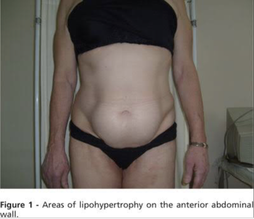

The background information here is that - this young lady with Type I Diabetes Mellitus presents with a 3cm swelling on the anterior surface of the hip.
Abnormal reaction in subcutaneous fat to insulin is called lipodystrophy which can be either hypertrophic or atrophic. Lipodystrophy is an exclusive complication of lean children and young Type 1 diabetic, although rarely can be seen in Type 2 diabetic.
Insulin lipohypertrophy denotes a benign tumor like swelling of fatty tissue at the injection site secondary to lipogenic effect of insulin, whereas lipoatrophy is considered an adverse immunological side effect of insulin therapy. Since the advent of recombinant human insulin and analogue, lipoatrophy, has virtually disappeared, whereas, lipohypertrophy still remains a serious local problem of insulin therapy.
With a swelling of 3cm, the best answer choice is development of hypertrophic lipodystrophy. Atrophic lipodystrophy will not have associated swelling, instead a depression should be observed.
Prevention, to avoid lipodystrophy, should take first place as there is little cure to it, and the best way is to educate the patient about rotating injection sites.
198. A 52-year-old woman has been suffering for 2 years from dull, occasionally exacerbating pain in her right subcostal area, occurring after eating high-fat foods, bitter taste in her mouth in the morning, constipations, and flatulence. Objectively she is obese, her body temperature is 36.9°C; there is a coating on the root of her tongue; the abdomen is moderately distended and painful in the area of gallbladder projection. What examination would be the most helpful for diagnosis making?
199. What should be prescribed as secondary prevention drugs for a patient with atrial fibrillation after an ischemic stroke caused by cardiac embolism?
200. A 55-year-old woman complains of thyroid gland enlargement that can be observed throughout the last 2 years and a discomfort during swallowing. Objectively she has signs of hypothyroidism. The thyroid gland on palpation is dense, non-fused with the surrounding tissues and mobile on swallowing. The regional lymph nodes are not enlarged. In the serum there are antithyroid antibodies detected. What is the most likely diagnosis?
201. A 19-year-old young man complains of cough with expectoration of purulent sputum in amount of 100 mL per day hemoptysis, dyspnea, increased body temperature up to 378°C, general weakness, weight loss. The patient\'s condition lasts for 4 years. Exacerbations occur 2-3 times a year. The patient presents with malnutrition, pale skin, cyanosis of the lips, drumstick (clubbed) fingers. Tympanic percussion sound in the lungs, weakened respiration, and various numerous moist crackles in the lower pulmonary segments on the left can be observed in this patient. In blood: erythrocytes - 3.2 • 1012/L,leukocytes -8.4 • 109/L,ESR- 56 mm/hour. On X- ray: lung fields are emphysematous, the left pulmonary root is deformed and dilated. What is the most likely diagnosis?
202. A 58-year old woman has type 2 diabetes mellitus that is compensated with diet and metformin. She prepares for cholecystectomy. Objectively, her height is 164 cm, weight is 90 kg, heart rate is 72/min., blood pressure is 130/80 mm Hg. Her abdomen is soft, painful in the right subcostal region. The liver is not enlarged. Fasting glucose - 6.2 mmol/L. Glycated hemoglobin - 6.5%. What further tactics of sugar-lowering therapy should be chosen in this case?
203. A 43-year-old man complains of facial edema, dyspnea, and difficult swallowing that appeared 3 hours after he was stung by an insect. Objectively the skin of his eyelids, cheeks, and lips is acutely hyperemic and edematous. What medicines should he be prescribed for emergency aid?
204. A 48-year-old woman was delivered into the surgical unit with wounds in her thigh. On examination the wound surface has a dirty-gray coating with unpleasant sweet smell. The wound content resembles a raspberry jelly. Skin tissues around the wound are glossy and turgid. Palpation reveals moderate crepitation in the tissues. What bacteria is the most likely to cause such inflammation?
205. A 26-year-old man complains of chills, rhinitis, dry cough, and fever up to 38°C. Examination shows him to be in a moderately severe condition; there are small pale pink non-merging spots on the skin of his back, abdomen, and extremities. Palpation reveals enlarged occipital and axillary lymph nodes. No information about vaccination history could be obtained. What is the likely etiology of this disease?
Explanation
Rubella ( accused by rubella virus) is an acute viral infection characterised by a short prodromal period, Exanthema ( rash) during three days and lymphadenopathy. The type of rash found is roseola and small macula-papula. Note that the rash elements do not merge ( non-merging spots) and are usually concentrated on the extensor surface of extremities, back, buttocks and outer surface of thigh.
Epstein-barr virus is one of the causative agents of Infectious mononucleosis- a disease characterised by prolonged fever, systemic lymphadenopathy,acute tonsillitis, acute adenoiditis, hepatosplenomegaly, along with typical blood changes such as lymphocytosis, monocytosis, presence of specific cells called atypical mononuclear cells or virocytes.
Mumps infection is a systemic disease characterised by hyperthermic syndrome,symmetrical or unilateral swelling of parotid glands, epididymo-orchitis etc.
206. A 38-year-old patient has been delivered by an ambulance to a surgical department with complaints of general weakness, indisposition, black stool. On examination the patient is pale,there are dotted hemorrhages on the skin of his torso and extremities. On digital investigation there are black feces on the glove. Blood test: Hb-108 g/L, thrombocytopenia. Anamnesis states that similar condition was observed 1 year ago. Make the diagnosis:
207. A 27-year-old man complains of pain in his leg joints, purulent discharge from the eyes, and painful burning sensations during urination. The disease onset was acute. He has a history of influenza. The patient smokes and drinks alcohol in excess. In his line of work he is often away on business trips. What is the most likely etiological factor of this disease?
208. A 57-year-old man, a miner, complains of a pain in his chest, dyspnea on physical exertion, excessive sweating, constant subfebrile tempeature, and cough that produces blood-streaked sputum. He has been smoking for approximately 40 years (2 packs a day) and frequently has “pneumonias”. Survey chest X-ray shows a triangular shadow in the middle lobe of the right lung. One of the apices of the shadow points to the lung root. Cardiac and mediastinal shadows are displaced toward the affected area. Make the provisional diagnosis:
209. A 31-year-old drug-addicted person complains of a cough with bloody expectorations, dyspnea, persistent fever, and leg edemas. The jugular veins are distended. There is a coarse pansystolic murmur detected above the base of the xiphoid process and in the second intercostal space on the left, close to the edge of the sternum. Heart sounds are clear; arrhythmia is detected, heart rate is 128/min., pulse - 82/min., blood pressure is 100/70 mm Hg. What is the most likely diagnosis?
210. A 17-year-old girl has been suffering from hepatic cirrhosis for 3 years. Lately her periods of excitation have been intermittent with depression, she does not sleep enough. Objectively her condition is severe, the girl is sluggish, gives one-word responses, has tremor in her extremities, her skin is icteric, with single hemorrhagic rashes. Name the likely complication of her disease:
211. A 34-year-old woman after rapidly changing her position from horizontal to vertical suddenly paled, fell down, her skin became moist, her limbs are cold, her pupils are dilated. The pulse is rapid and thready blood pressure is 50/25 mmHg. What condition has likely developed in the patient?
212. A woman complains of a severe pain in her throat on the left,difficult swallowing and mouth opening, elevated body temperature, and general malaise. The onset of the disease was 4 days ago after a case of tonsillitis. Examination detects a trismus of the masticatory muscles, the left tonsil is displaced toward the midline, the anterior palatal arch is infiltrated and protrudiiig. The regional lymph nodes on the right are enlarged and painful on palpation. Make the diagnosis:
213. During winter epidemics of influenza caused predominantly by virus A/California/04/2009 (H1N1), on the 2nd day after the disease onset a 30-year-old hospitalized man presented with high fever, dry cough, myalgia, headache, and general weakness. What should be prescribed as an etiotropic treatment in this case?
214. A 30-year-old woman complains of itching skin, predominantly in the evening and at night. The condition lasts for 2 weeks already On the skin of the interdigital folds, mammary glands, abdomen, buttocks, and thighs there are numerous fine papular and papulovesicular rashes located in pairs, excoriations. There is no rash on the face and neck. Similar rash is observed in the husband of the patient. What is the most likely diagnosis?
Explanation
Scabies is a parasitic skin infestation caused by the Sarcoptes scabiei. It is characterised by intense pruritus (itching) at night; this characteristic and the location of the rashes helps us to diagnose scabies. The primary lessios found are usually papules, vesicles or burrows ( accompanied by excoriations and pustules) which are commonly seen in interdigital folds, flexor surfaces of the wrist, axillary folds, buttocks (closed areas of the body). Additionally in children, elderly persons, and immunosuppressed patients: scalp, face, neck, under the nail, palms of hands, and soles of feet.
Neurodermatitis is also referred to as Lichen simplex chronicus; it is chracterised by chronic itching and scaling localised on the wrist, neck, forearm, legs and anal region.
Epidermophytosis is referred to as ‘Athlete’s foot’. 215. A 20-year-old patient complains of a severe headache, double vision, weakness, fever; irritability Objectively: body temperature is 38.1°C, the patient is reluctant to contact, sensitive to stimuli. There are ptosis of the left eyelid, exotropia, anisocoria S > D, and pronounced meningeal syndrome. On lumbar puncture the cerebrospinal fluid flowed out under a pressure of 300 mmHg, the fluid is clear, slightly opalescent. 24 hours later there appeared a fibrin film. Protein - 1.4 g/L, lymphocytes - 600 per mm3, glucose - 0.3 mmol/L. What is the provisional diagnosis?
216. A 75-year-old man in a severe condition suffers from dyspnea at rest, marked weakness, and arrhythmia. Abdominal aortic pulsation is observed, further on there is a systolic murmur detected. Palpation reveals a volumetric formation in the mesogastrium. Blood pressure is 70/40 mm Hg. There is no pulsation over the femoral arteries. Oliguria is detected. Which diagnosis is the correct one?
217. A dweller of the northern Dnieper area, a fisherman, for the last several days has been complaining of a discomfort in his right subcostal region, periodical episodes of diarrhea, intermittent with constipations, frequent skin rashes. Abdominal ultrasound shows enlarged liver and pancreatic head. Make the provisional diagnosis:
218. An 18-year-old patient complains of skin rash. The patient has been suffering from this condition for 5 years. The first instance of this disease occurred after a car accident. Objectively: the patient presents with a papular rash covered in silvery scales, “thimble” sign (small pits on the nails), affected joints. What is the most likely diagnosis?
219. During regular medical examination a lyceum student presents with signs of cheilitis that manifests as epithelial maceration in the area of lip seal. The lips are bright-red, with single vertical cracks covered with brown-red scabs. These clinical signs are most likely caused by insufficient content of the following in the diet:
220. A 72-year-old man with pneumonia complains of marked dyspnea, chest pain, severe cough with expectoration, t° is 39.5-40°C, no urination for the last 24 hours. Objectively the patient is conscious. Respiratory rate is 36/min. Percussion sound is dull over the right lower pulmonary lobe; on auscultation, there is bronchial respiration and numerous moist crackles. Blood pressure is 80/60 mm Hg, Heart rate is 120/min. Heart sounds are muffled, there is tachycardia. What tactics should the family doctor choose for the management of this patient?
221. A 48-year-old man came to a doctor with complaints of vomiting that brings no relief and a burning pain in his left subcostal region that irradiates to the left lumbar region. These signs appeared after a meal. The Ortner’s and Mayo-Robson’s signs are positive. In the blood: leukocytosis and increased ESR. In the urine: elevated diastase levels. Make the diagnosis:
222. A 32-year-old woman complains of general weakness, low-grade fever persisting for 4 months, lumbar pain, and dysuria. Anamnesis includes frequent acute respiratory diseases, overexposure to cold, low-calorie diet, a case of pulmonary tuberculosis in childhood. Clinical urine analysis: pH- 4.8, leukocyturia, hematuria. Complete blood count: leukocytosis, lymphocytosis, raised ESR. Urography concludes: dilatation of renal pelvis and calyceal system of both kidneys, foci of calcification in the projection of right kidney parenchyma. What is the most likely diagnosis?
223. An unconscious patient was delivered to a hospital by an ambulance. Objectively his body temperature is 39°C, he presents with convulsions and red dry skin. It is known that the patient works as a stoker in the boiler room. What is the likely diagnosis?
Explanation
From anamnesis, we observe that this patient works in a very hot/ steaming environment; the signs shown by this patient i.e., a red dry skin and a temperature of 39℃ indicates the effect of heat on the patient. Heat stroke is a form of hyperthermia in which the body temperature is elevated dramatically. The cause of heat stroke is an elevation in body temperature, often accompanied by dehydration.
The most common symptoms of CO (carbon monoxide) poisoning are headache, dizziness, weakness, upset stomach, vomiting, chest pain, and confusion on the background of inhalation of fumes from cars, generators etc.
224. 2 hours after eating unknown mushrooms, a 28-year-old man sensed a decrease in his mobility and deterioration of his ability to focus. This condition was then followed by a state of agitation and agression. On examiantion he is disoriented and his speech is illegible. 4 hours later he developed fetor hepaticus and lost his consciousness. What syndrome can be observed in this patient?
225. A 19-year-old girl complains of moderate itching and hair loss on her head. Objectively on the skin of her occipital region there is a single round erythematous focus 3 cm in diameter with clear margins. Asbestos-like scales can be observed on the surface of the lesion. The hair in the focus of the lesion is broken off at the length of 6-8 mm. What is the most likely diagnosis?
226. A woman complains of frequent watery stool (up to 9-10 times per day) with mucus and blood admixtures, dull pain in the hypogastrium, weight loss of 4 kg within the last year. Objectively: malnutrition, dry skin, low turgor, aphthous stomatitis. The abdomen is soft, the sigmoid colon is spastic and painful on palpation. Occult blood test is positive. Fibrocolonoscopy: edema, hyperemia, mucosal granulation, pseudopolyp, small ulcers with irregular edges. Make the diagnosis:
Explanation
Differences between Ulcerative Colitis and Crohn Disease
|
Ulcerative Colitis
|
Crohn Disease
|
|
Only colon involved
|
Panintestinal
|
|
Continuous inflammation extending proximally
from rectum
|
Skip-lesions with intervening normal mucosa
|
|
Inflammation in mucosa and submucosa only
|
Transmural inflammation
|
|
No granulomas
|
Noncaseating granulomas
|
|
Perinuclear ANCA (pANCA) positive
|
ASCA positive
|
|
Bleeding (common)
|
Bleeding (uncommon)
|
|
Fistulae (rare)
|
Fistulae (common)
|
Cathartic colon
Occult blood in stool is indicative of a hemorrhagic process from the colon, while the results from the colonoscopy indicates the pathology is located in the colon.
Irritable bowel syndrome is an inflammatory bowel disease characterised by recurrent abdominal cramps (pain), change in form (consistency) and frequency of stool, constipation etc. . This condition is common in middle aged women and is associated with underlying conditions such as stress, anxiety, depression or a previous case of intestinal infection.
227. A patient was delivered to a surgical department after a road accident with a closed trauma of chest and right-sided rib fracture. The patient was diagnosed with right sided pneumothorax; it is indicated to perform drainage of pleural cavity. Pleural puncture should be made:
228. An 18-year-old patient was admitted to a hospital with complaints of headache, weakness, high fever, sore throat. Objectively: enlargement of all groups of lymph nodes was revealed. The liver is enlarged by 3 cm, spleen- by 1 cm. In blood: leukocytosis, atypical lymphocytes - 15%. What is the most probable diagnosis?
229. A 45-year-old patient complains of pain in the epigastric region, left subcostal area, abdominal distension, diarrhea, loss of weight. He has been suffering from this condition for 5 years. Objectively: tongue is moist with white coating near the root; deep palpation of abdomen reveals slight pain in the epigastric region and Мауо-Robson’s point. Liver is painless and protrudes 1 cm from the costal arch. Spleen cannot be palpated. What disease can be primarily suspected?
230. A 25-year-old patient was delivered to an infectious diseases unit on the 3rd day of illness with complaints of headache, pain in lumbar spine and gastrocnemius muscles, high fever, chill. Objectively: condition of moderate severity. Scleras are icteric. Pharynx is hyperemic. Tongue is dry with dry brown coating. Abdomen is distended. Liver is enlarged by 2 cm. Spleen is not enlarged. Palpation of muscles, especially gastrocnemius muscles, is painful. Urine is dark in colour. Stool is normal in colour. The most likely diagnosis is:
231. A 47-year-old patient came to see a doctor on the 7th day of disease. The disease is acute: after the chill body temperature rose to 40*C and lasted up to 7 hours, then dropped abruptly, which caused profuse sweat. There were three such attacks occurring once every other day. Two days ago the patient arrived from Africa. Objectively: pale skin, subicteric sclera, significantly enlarged liver and spleen. What is the cause of fever attacks in this disease?
232. A 24-year-old patient complains of general weakness, dizziness, body temperature rise up to 37,5*C, sore throat, neck edema, enlargement of submaxillary lymph nodes. Objectively: mucous membrane of oropharynx is edematic and cyanotic, tonsils are enlarged and covered with films that spread beyond the tonsils and cannot be easily removed. What is the leading mechanism of this illness development?
233. A patient’s X-ray image (anteroposterior projection) shows deformation of lung pattern, pneumofibrosis, reticular (honeycomb) lung pattern of lower pulmonary segments, cylindric and fusiform lumps. The most likely diagnosis is:
234. A 58-year-old patient complains of general weakness, weight loss up to 10 kg within the last 1,5 months, progressive pain in the small of the back, raise of blood pressure to 220/160mm Hg, subfebrile body temperature. Objectively: tuberous slightly movable lump can be palpated in the right subcostal area; veins of spermatic cord and scrotum are dilated. Blood test: Hb - 86 g/l, ESR - 44 mm/h. Urine test: specific gravity - 1020, proteine - 0,99g/l, erythrocytes - all field of vision, leukocytes- 4-6 in the field of vision. The provisional diagnosis is:
235. A 37-year-old patient complains of pain in the lumbar spine, which increases during walking, limited mobility, edema of the right side of abdomen. Focal tuberculosis is recorded in the anamnesis. X-ray shows destruction of adjacent surfaces of the bodies of the 1-2 lumbar vertebrae, vertebral bodies height is decreased, intervertebral fissure cannot be detected. Abdominal US reveals a growth 15x20 cm in size in the retroperitoneal space and echo signs of liquid. What diagnosis can be suspected?
236. A 53-year-old woman complains of weight loss up to 10 kg within the last 2 years, liquid foul smelling stool two times a day that poorly washes off the toilet, periodic bouts of nausea, girdle pain in the upper abdomen. Objectively: pain in Gubergrits zone (on the right from navel) and Mayo Robson’s point. Biochemical blood analysis: glucose - 3,2 mmol/l, bilirubin -16,5 micromole/l, crude protein - 56,4 g/l. Urine diastase - 426 g/h/l. D-xylose test (oral administration of 25 g of dxylose) after 5 hours reveals 3 g of xylose in urine. The most likely diagnosis is:
237. A 23-year-old patient after intake of brake fluid has developed anuria that has been lasting for 5 days already. Creatinine level increased up to 0,769 mmol/l. What treatment tactics should be chosen in the given case?
238. A patient complains of weight gain, chill, edemas, xeroderma, somnolence, difficulties with focusing. Objectively: height is 165 cm; weight is 90 kg; body proportions are of female type, to - 35,8*C, heart rate - 58/min, BP -105/60 mm Hg. Heart sounds are weakened, bradycardia is observed. Other internal organs have no changes. Thyroid gland cannot be palpated. Milk secretion from mammary glands is observed. Hormone investigation revealed increased levels of thyroid stimulating hormone (TSH) and prolactin, and decreased level of thyroxine (T4). Which one is the cause for obesity?
239. A 54-year-old patient complains of weakness, jaundice, itching skin. Disease onset was 1,5 months ago: fever up to 39*C appeared at first, with progressive jaundice developed 2 weeks later. On hospitalisation jaundice was severely progressed. Liver cannot be palpated. Gallbladder is enlarged and painless. Blood bilirubin is 190 micromole/l (accounting for direct bilirubin). Stool is acholic. What is the most likely jaundice genesis in this patient?
240. A 40-year-old patient complains of constant moderate pain in the lumbar spine and significantly reduced mobility. The patient has been suffering from this condition for the last 7 years since pain appeared first in the sacrum area. X-ray: ankylosis of sacroiliac articulation, significant narrowing of intervertebral joint fissures of lumbar vertebrae and calcification of spinal ligaments. What pathology is most likely to cause such X-ray image?
241. After overexposure to cold a 45-year- old woman developed acute pain in her suprapubic and lumbar areas during urination, sharp pains at the end of urination, false urges to urinate. Urine is turbid with blood streaks. The doctor suspects urinary tract infection. What results of laboratory analysis would be the most indicative of such infection?
Explanation
Urinary tract infections are commonly associated with dysuria, urinary frequency, urinary urgency and lumboabdominal pains.
When you have a UTI, the lining of the bladder and urethra become inflammed and irritated just as your throat does when you have phrayngitis, common cold - upper respiratory tract infections. The irritation can cause pain in your lower abdomen - pelvic area and even lower back, and will usually make you feel like urinating more often (false urges). Burning or pain when urinating is the most common symptom. You may even feel a strong urge or need to urinate but only get a few drops. This is because the bladder is so irritated that it makes you feel like you have to urinate, even when you don't have much urine in your bladder. UTIs can be found by analyzing a urine sample for Urine Microscopy/Culture/Sensitivity (Urine M/C/S). The urine is examined under a microscope for bacteria or white blood cells (leukocyturia), which are signs of infection. Blood can also be found in the urine sample (hematuria); this can be due to an infective or mechanical cause.
The other options given are more specific for kidney pathologies - proteinuria, gross hematuria, creatinine and urea. Rarely are they elevated in pathologies relating to Urinary Tract Infections except in cases where UTI’s become complicated and the kidney is affected after a prolonged and untreated UTI.
242. A 34-year-old man complains of pale edema of the face, feet, shins, and lumbar area, elevated blood pressure up to 160/100 mm Hg, and general weakness. He has a clinical history of nonspecific ulcerative colitis. Objectively: pulse - 84/min., rhythmic, blood pressure - 165/100 mm Hg; edemas all over the body; the skin is pale and dry, with low turgor. The kidneys cannot be palpated, on an attempt to palpate them they are painless. Blood test: erythrocytes - 3.0∙1012/L, Hb- 100 g/L, erythrocyte sedimentation rate - 50 mm/hour. Urinalysis: proteins - 3.5 g/L, erythrocytes -7-10 in the vision field, leukocytes - 5-6 in the vision field. Daily proteinuria 6 grams. What analysis should be conducted additionally to verify the diagnosis?
243. A 40-year-old man with Bekhterev disease (ankylosing spondylitis) complains of elevated body temperature up to 37.8°C, back pain and stiffness, especially observed during the second half of the night. This condition has been lasting for 2 years. Objectively: reduced spinal mobility, painful sacroiliac joint, erythrocyte sedimentation rate - 45 mm/hour. X-ray shows narrowing of the intervertebral disc space and of the sacroiliac joint. What eye pathology is often associated with this type of disease progression?
244. A 27-year-old man complains of pain in his leg joints, purulent discharge from the eyes, and painful burning sensations during urination. Disease onset was acute. He has a history of influenza. The patient smokes and drinks alcohol in excess. In his line of work he is often away on business trips. What is the most likely etiological factor of this disease?
Explanation
Reactive arthritis is an inflammatory arthritis which manifests after several days to weeks after a gastrointestinal or genitourinary infection. It is also described as a classic triad of arthritis, urethritis and conjunctivitis. However, a majority of patients do not present with the classic triad. It was previously called "Reiter syndrome."
Classic triad:
Conjunctivitis: purulent discharge from the eyes
Urethritis: painful burning sensations during urination
Arthritis: pain in leg joints
Reactive arthritis is known to be triggered by a bacterial infection, particularly of the genitourinary (Chlamydia trachomatis, Neisseria gonorrhoeae, Mycoplasma hominis, and Ureaplasma urealyticum) or gastrointestinal (GI) tract (Salmonella enteritidis, Shigella flexneri, and dysenteriae, Yersinia enterocolitica, Campylobacter jejuni, Clostridium difficile).
The classic triad can be remembered using this:
Conjunctivitis; Urethritis; Arthritis - Can’t See; can’t pee, can’t bend my knee
245. A 51-year-old man complains of vomiting with blood. He has been drinking alcohol excessively. Health disorder has been observed since he was 40, when he first developed jaundice. On examination the skin and visible mucosa are icteric, with a stellate vascular pattern. The patient is malnourished and presents with abdominal distension, umbilical hernia, and ascites. The edge of the liver is tapered and painless, +3 cm, the spleen is +2 cm. Blood test: Hb - 80 g/L, leukocytes -3∙109/L, platelets - 85∙109/L. What is the cause of portal hypertension in this patient?
Explanation
Cirrhosis is a complication of liver disease that involves loss of liver cells and irreversible scarring of the liver. The scarring is accompanied by loss of functioning liver cells. Cirrhosis is usually a late-stage disease of the liver that is not reversible in severe cases.
Hepatitis B, C, and chronic alcohol abuse are frequent causes (Question stem - patient takes alcohol excessively). Initially patients may experience fatigue, weakness and weight loss. During later stages, patients may develop jaundice (yellowing of the skin), gastrointestinal bleeding, abdominal swelling and confusion.
The most common cause of portal hypertension is cirrhosis of the liver. Portal hypertension is an increase in the blood pressure within a system of veins called the portal venous system. Veins coming from the stomach, intestine, spleen, and pancreas merge into the portal vein, which then branches into smaller vessels and travels through the liver. If the vessels in the liver are blocked due to liver damage, blood cannot flow properly through the liver. As a result, high pressure in the portal system develops.
Budd-Chiari syndrome is a condition in which the hepatic veins (veins that drain the liver) are blocked or narrowed by a clot (mass of blood cells). It may lead to portal hypertension, ascites, cirrhosis etc. Budd-Chiari syndrome can be caused by conditions and situations that cause your blood to clot (form a blockage) like sickle cell disease, pregnancy, myeloproliferative disorders. It is not caused by excessive alcohol intake or Hepatitis.
Hemochromatosis, Constrictive Pericarditis and Thrombosis of the splenic vein will not directly lead to portal hypertension.
246. A 53-year-old man complains of general weakness, loss of appetite, and painful vesicles appearing on his skin. The disease onset occurred suddenly, after hyperinsolation one week ago. Examination detects isolated vesicles with wrinkled opercula and occasional painful erosions on the skin of the patient’s torso and limbs. Nikolsky sign is positive. What is the most likely diagnosis?
Explanation
Nikolsky's sign has been a very useful diagnostic tool in cases of skin disorders like pemphigus, toxic epidermal necrolysis, etc., The sign is demonstrated when lateral pressure is applied on the border of an intact blister, which results in the dislodgment of the normal epidermis and extension of the blister. For the Nikolsky sign in patients with active blistering, firm sliding pressure with a finger separates normal-appearing epidermis, producing an erosion. This sign is made possible due to the loss of cell-to-cell adhesion in the epidermis.
Acantholysis is the loss of intercellular connections, such as desmosomes, resulting in loss of cohesion between keratinocytes, seen in diseases such as pemphigus vulgaris. It is absent in bullous pemphigoid, making it useful for differential diagnosis. Hence, Nikolsky’s sign will be negative in non-acantholytic pemphigus.
Nikolsky's sign is pathognomonic for pemphigus, toxic epidermal necrolysis, and staphylococcal scalded skin syndrome (SSSS). This sign basically differentiates intraepidermal blisters from subepidermal blisters.
Toxicoderma are usually caused by drugs and this patient has no history of taking any medication that would have resulted in toxicoderma. Dermatitis herpetiformis is an autoimmune blistering disorder associated with a gluten-sensitive enteropathy (patient wasn’t diagnosed of celiac disease). Herpes usually presents as grouped vesicles, meanwhile, on examination of the patient isolated vesicles were detected.
247. A 48-year-old woman developed insomnia, depressive mood, anxiety, fears and suicidal thoughts after the death of her husband that occurred one month ago. During her stay in the hospital she speaks in a low voice, is depressed, anxious, avoids sleeping, refuses to eat. What medications should be prescribed in this case?
248. A woman has been provisionally diagnosed with pheochromocytoma. At the stage of intermission her BP is within norm; there is a tendency towards tachycardia. No urine pathologies. The decision has been made to perform a provocative test with histamine. What drug should be kept close at hand for emergency aid in case of positive test result?
249. A 52-year-old man for the last 3 years has been suffering from difficult swallowing of solid food, burning retrosternal pain that aggravated during eating, loss of body mass, and occasional vomiting with undigested food. Esophageal X-ray shows S-shaped deformation of the esophagus and its dilation; at the cardiac orifice the esophagus is constricted; esophageal mucosa is smooth, without signs of peristalsis. Make the provisional diagnosis:
250. A 72-year-old man diagnosed with ischemic heart disease presents with diffuse cardiosclerosis, permanent tachysystolic atrial fibrillation, heart failure IIa, FC III. Objective examination of vital signs: blood pressure is 135/80 mm Hg, heart rate is 160/min., pulse is 125/min. Left ventricular ejection fraction is 32%. What drug is indicated in this case and should be prescribed to the patient?
251. A 32-year-old woman complains of tumor-like formation on the anterior surface of her neck that appeared 2 years ago. Within the last 3 months the tumor has been rapidly growing. It hinders swallowing and impairs speech; the tumor causes a sensation of pressure. Objectively the skin moisture is normal, pulse is 80/min., rhythmic, blood pressure is 130/80 mm Hg. In the right lobe of the thyroid gland there is a dense lumpy node 3.0x3.5 cm that moves during swallowing. Scanning image shows a ”cold nodule” in the thyroid gland. Make the provisional diagnosis:
252. A 32-year-old woman complains of body weight loss despite her increased appetite, nervousness, and tremor of the extremities. Objectively: the skin is moist; the thyroid gland is diffusely enlarged, painless, soft, and mobile. Blood test: increased level of T3, T4, and thyroid-stimulating hormone (TSH). What is the most likely diagnosis?
Explanation
Diffuse toxic goiter or grave’s disease is an autoimmune disease characterised by an increased amount of thyroid hormones ( T3- triiodothyronine and T4- thyroxine). Symptoms include; loss of weight, with increased appetite, increased sweating, cardiac findings (tachycardia, increased risk for atrial fibrillation), tremors of extremities, heat intolerance, diarrhea, anxiety etc.
It is the most common cause of hyperthyroidism and more common in females than males. There is production of IgG antibodies against the TSH receptor (stimulating type of antibody - Type II hypersensitivity reaction)
Hashimoto’s thyroiditis is a destructive autoimmune thyroiditis leading to hypothyroidism.
In Diffuse non-toxic goitre, thyroid function is normal and patients are often asymptomatic. It manifests without hyperthyroidism, hypothyroidism, or inflammation. 253. A 45-year-old man, a farmer, presents with acute onset of a disease. He complains of headache, high temperature, pain in the gastrocnemius muscles, icteric face, and dark urine. Objectively: body temperature - 38°C, blood pressure - 100/70 mm Hg, conjunctival hemorrhages, hepatosplenomegaly, and oliguria. What is the most likely provisional diagnosis?
Explanation
Leptospirosis is an acute generalized infectious disease, characterized by extensive vasculitis, caused by spirochetes of the genus Leptospira. It presents with flu-like symptoms, myalgias (majorly calf muscles eg gastrocnemius), jaundice (icteric face), Photophobia etc. The icterohemorrhagic form of leptospirosis presents with a severe form of jaundice and azotemia from the liver and kidney dysfunction coupled with anemia. Leptospira is commonly found in water contaminated with animal urine.
N/B Virus hepatitis has a gradual onset, without chills, the temperature rises at the pre-icteric period. Muscle pains, scleritis, conjunctivitis are not characteristic of it.
Brucellosis is caused by gram negative bacteria, transmitted via ingestion of contaminated animal products (eg, unpasteurized milk). Typically presents with fever, night sweats, and arthralgia but jaundice is usually absent.
254. A 38-year-old patient has been brought by an ambulance to the surgical department with complaints of general weakness, indisposition, black stool. On examination the patient is pale, there are dotted hemorrhages on the skin of his torso and extremities. On digital investigation there are black feces on the glove. Blood test: Hb - 108 g/L, thrombocytopenia. Anamnesis states that a similar condition was observed 1 year ago. Make the diagnosis:
255. A 42-year-old man, a worker at the meat processing factory, developed an itching spot on his lower jaw, which gradually transformed into a slightly painful carbuncle 3 cm in diameter, surrounded by a painless swelling that reaches the clavicle. Temperature is subfebrile, under 37.8°C. The doctor suspects anthrax. What drug should this man be prescribed for treatment?
Explanation
Bacillus anthracis is an aerobic, non-motile, spore forming, large non-hemolytic Gram-positive rod that grows well on blood agar. The clinical manifestations of human anthrax - Cutaneous and Inhalation.
Cutaneous anthrax, the most common form of naturally occurring disease, begins as a small, painless, pruritic papule that within 2 days enlarges, develops vesicles, and ulcerates to form an eschar. Inhalational anthrax follows inhalation of infectious doses of anthrax spores.
Four antibiotics are approved for use for post exposure prophylaxis following exposure to aerosolized spores of B. anthracis: doxycycline, ciprofloxacin, levofloxacin, and parenteral procaine penicillin G. Of all these four antibiotics, only Penicillin is listed as a choice for this question. 256. A patient is being treated in the tuberculosis clinic. Throughout the last 3 weeks he has been suffering from headaches of increasing intensity. Neurological examination detects nuchal rigidity without focal signs. Make the provisional diagnosis:
257. A 37-year-old man suddenly developed acute headache accompanied by nausea, vomiting, and impaired consciousness. Objectively blood pressure is 190/120 mm Hg, the face is hyperemic. Patient’s consciousness is clouded, his answers to the questions are short, monosyllabic. Movement and sensory disturbances are absent. Meningeal signs are positive. Cerebrospinal fluid contains blood. What provisional diagnosis can be made?
258. A 20-year-old student after failing an exam developed complaints of a sensation of a round foreign body in her throat, difficult swallowing. She fixates on her condition, limits her diet, often cries, seeks attention, exhibits demonstrative attitude. She is highly susceptible to psychotherapeutic suggestion. What psychiatric diagnosis can be made in this case?
259. A 39-year-old man suffers from chronic rheumatic heart disease. He complains of dyspnea during physical exertion, cough with expectoration, and palpitations. Ausculation detects intensified I heart sound and diastolic murmur; the sound of opening mitral valve can be auscultated at the cardiac apex. The II heart sound is accentuated over the pulmonary artery. The patient is cyanotic. X-ray shows dilated pulmonary root and enlargement of the right ventricle and left atrium. What is the most likely diagnosis?
Explanation
|
Valvular diseases
|
Mitral stenosis
|
Aortic stenosis
|
Aortic regurgitation
|
Mitral regurgitation
|
Tricuspid regurgitation
|
|
Main causes and risk factors
|
Almost always caused by rheumatic heart disease
|
-Calcification of tricuspid aortic valve with age
(>50%)
-Calcification of bicuspid aortic valve
(30-40%)
-Rheumatic fever
(<10%)
Hypertension, diabetes mellitus, hyperlipoproteinemia and uremia may speed up the process.
|
Acute
-Infective endocarditis
-Trauma
Chronic
-Primary valvular: rheumatic fever, bicuspid aortic valve, Marfan's syndrome, Ehlers–Danlos syndrome, ankylosing spondylitis, systemic lupus erythematosus
-Disease of the aortic root: syphilitic aortitis, osteogenesis imperfecta, aortic dissection, Behçet's disease, reactive arthritis, systemic hypertension
|
Acute
-Endocarditis, mainly S. aureus
-Papillary muscle rupture or dysfunction, including mitral valve prolapse
Chronic
-Rheumatic fever
-Marfan's syndrome
-Cardiomyopathy
|
Usually secondary to right ventricular dilation
Other causes: Tricuspid endocarditis, rheumatic fever, Ebstein's anomaly, carcinoid syndrome and myxomatous degeneration
|
|
Symptoms
|
Heart failure symptoms, such as dyspnea on exertion, orthopnea and paroxysmal nocturnal dyspnea
-Palpitations
-Chest pain
-Hemoptysis
-Thromboembolism
-Ascites and edema (if right-sided heart failure develops)
Symptoms increase with exercise and pregnancy
|
-Heart failure symptoms, such as dyspnea on exertion (most frequent symptom), orthopnea and paroxysmal nocturnal dyspnea
-Angina pectoris
-Syncope, usually exertional
|
-Heart failure symptoms, such as dyspnea on exertion, orthopnea and paroxysmal nocturnal dyspnea
Palpitations,
-Angina pectoris
-In acute cases: cyanosis and circulatory shock
|
-Heart failure symptoms, such as dyspnea on exertion, orthopnea and paroxysmal nocturnal dyspnea
-Palpitations
-Pulmonary edema
|
Symptoms of right-sided heart failure, such as ascites, hepatomegaly, edema and jugular venous distension
|
|
Medical signs
|
-Opening snap followed by a low-pitched diastolic rumble with presystolic accentuation.
The opening snap follows closer to the S2 heart tone with worsening stenosis.
The murmur is heard best with the bell of the stethoscope lying on the left side and its duration increases with worsening disease.
-Loud S1 - may be the most prominent sign
-Advanced disease may present with signs of right-sided heart failure such as parasternal heave, jugular venous distension, hepatomegaly, ascites and/or pulmonary hypertension (presenting with a loud P2.
Signs increase with exercise and pregnancy
|
Systolic murmur of a harsh crescendo-decrescendo type, heard in 2nd right intercostal space, radiating to the carotid arteries
-Pulsus parvus et tardus, that is, diminished and delayed carotid pulse
-Fourth heart sound
-Decreased A2 sound
-Sustained apex beat
Precordial thrill
|
-Increased pulse pressure by increased systolic and decreased diastolic blood pressure, but may not be significant if acute
-Diastolic decrescendo murmur best heard at left sternal border
Water hammer pulse
-Austin Flint murmur
-Apex beat displaced down and to the left
-Third heart sound may be present
|
Holosystolic murmur at the apex, radiating to the back or clavicular area
Commonly atrial fibrillation
Third heart sound
Laterally displaced apex beat, often with heave
Loud, palpable P2, heard best when lying on the left side
|
-Pulsatile liver
-Prominent V waves and rapid descents in jugular venous pressure
-Inspiratory third heart sound at left lower sternal border (LLSB)
-Blowing holosystolic murmur at LLSB, intensifying with inspiration, and decreasing with expiration and -Valsalva maneuver
-Parasternal heave along LLSB
-Atrial fibrillation is usually present
|