NOTEPAD
Results
of 150 questions answered correctly

You have reached of 150 points, ( %)

Your time

Question 1 of 150

1.  

A patient was brought by an ambulance to the impatient department. He was provisionally diagnosed with acute pancreatitis. To confirm this diagnosis, it is necessary to measure the activity of a certain enzyme in the patient’s blood and urine. Name the enzyme:

Explanation

Serum amylase remains the most commonly used biochemical marker for the diagnosis of acute pancreatitis. Acute pancreatitis typically presents with severe, constant upper abdominal pain which may radiate through to the back and be associated with nausea and vomiting. As atypical presentations are frequent and there is a wide differential diagnosis, confirmatory tests are required to confirm the diagnosis of acute pancreatitis. Three enzymes derived from pancreatic acinar cells - amylase, lipase, and the proenzyme trypsinogen - have been tested as biochemical markers of acute pancreatitis; serum amylase is the most commonly used of these in clinical practice.

Aspartate aminotransferase (AST) and alanine aminotransferase (ALT) are enzymes found mainly in the liver, but also found in red blood cells, heart cells, muscle tissue and other organs. The levels of AST and ALT in serum can help people diagnose body tissue pathology especially the liver and the heart. 


Cholinesterase is an enzyme that breaks down the neurotransmitter Acetylcholine. In medicine, Lactate dehydrogenase (LDH) is commonly used as a marker of tissue breakdown as LDH is abundant in red blood cells and can function as a marker for hemolysis.

2.

Blood test of the patient revealed albumin content of 20 g/L and increased activity of lactate dehydrogenase isoenzyme 5(LDH5). These results indicate disorder of the following organ.

Explanation

The two serum proteins measured to assess liver function are albumin and globulin. Albumin, produced only in the liver, is the major plasma protein that circulates in the bloodstream.

Albumin: 35 - 50g/L norm (So, an Albumin level of 20g/L is significantly decreased).

Albumin is synthesized in the liver, and low serum albumin can be indicative of liver failure or diseases such as cirrhosis and chronic hepatitis. A low serum albumin indicates poor liver function. Decreased serum albumin levels are not seen in acute liver failure because it takes several weeks of impaired albumin production before the serum albumin level drops. The most common reason for a low albumin is chronic liver failure caused by cirrhosis. 

There are five types of LDH. They are known as isoenzymes. The five isoenzymes are found in different amounts in tissues throughout the body.

  • LDH-1: found in heart and red blood cells

  • LDH-2: found in white blood cells. It is also found in heart and red blood cells, but in lesser amounts than LDH-1.

  • LDH-3: found in lung tissue

  • LDH-4: found in white blood cells, kidney and pancreas cells, and lymph nodes

  • LDH-5: found in the liver and muscles of skeleton


When tissues are damaged or diseased, they release LDH isoenzymes into the bloodstream.

3.

A 25-year-old woman complains of deteriorating vision. Examination revealed a defect in accommodation, the pupil is dilated and unresponsive to light. What muscles are functionally disturbed in this case?

Explanation

From the question stem, you will notice that the patient has a defect in accommodation and the pupil is dilated (which means it doesn't constrict).

Accommodation is determined by the ciliary muscle, which is a circular (constrictor) smooth muscle that is attached to the lens by suspensory ligaments. The ciliary muscle has only parasympathetic innervation (mediated by acetylcholine acting through muscarinic receptors).

The iris sphincter muscle, also known as the pupillary sphincter or sphincter pupillae, is a muscle located in the colored part of the eye called the iris. The sphincter muscle fibers are located near the pupillary margin and are slightly anterior to the pigmented epithelium of the iris. It encircles the pupil of the iris and functions to constrict the pupil in bright light via the pupillary light reflex or during accommodation. By controlling the diameter of the pupil, the iris controls the amount of light that reaches the back of the eye at the retina.

The question clearly states that there is a defect in accommodation which is clearly a function of the ciliary muscle and the pupil is dilated which means the pupil does not constrict hence, this is a pathology of the Iris sphincter.

The ciliary muscle, Iris sphincter and Iris dilator muscles are the intrinsic muscles of the eye. They are involved in accommodation (ciliary muscle); dilating (Iris dilator/Dilator pupillae) and constricting the eye (Iris sphincter/Sphincter pupillae).


Extrinsic muscles of the eye: Superior oblique; Inferior oblique; Lateral rectus; Medial rectus; Superior rectus and Inferior rectus muscles. They are involved in moving the eyeball up, down, left and right.

Ciliary Muscle and Iris Sphincter

4.

One of the causes of pernicious anemia is the disturbed synthesis of transcorrin – Castle’s intrinsic factor in the parietal cells of the stomach. What substance is called Castle’s extrinsic factor?

Explanation

The term intrinsic factor was coined in the late 1920s by the American physician William B. Castle, whose research into the cause of pernicious anemia indicated that two substances were involved: one that is produced in the body (intrinsic) and the other—an extrinsic factor, later identified as vitamin B12—that is supplied in the diet.

Intrinsic factor, a glycoprotein with which vitamin B12 (cyanocobalamin) must combine to be absorbed by the gut. Intrinsic factor is secreted by parietal cells of the gastric glands in the stomach, where it binds with the vitamin. Thus bound, intrinsic factor protects vitamin B12 from digestion as it passes through the gastrointestinal tract and facilitates the vitamin’s absorption in the ileum of the small intestine. Vitamin B12 is necessary for red blood cell maturation. A lack of intrinsic factor may result in inadequate absorption of the vitamin and cause pernicious anemia. Vitamin B12 and Intrinsic Factor
5.

Proliferation of connective tissue in the liver parenchyma (fibrosis) that results from chronic disorders causes of blood flow disturbance in the classical hepatic lobules. Describe the blood flow direction in these lobules:

Explanation

In the hepatic portal system, the liver receives a dual blood supply from the hepatic portal vein and hepatic arteries. The hepatic portal vein carries venous blood drained from the spleen, gastrointestinal tract and its associated organs; it supplies approximately 75% of the liver’s blood. The hepatic arteries supply arterial blood to the liver and account for the remainder of its blood flow.

Oxygen is provided from both sources; approximately half of the liver’s oxygen demand is met by the hepatic portal vein, and half is met by the hepatic arteries. Blood flows through the liver tissue and empties into the central vein of each lobule. The central veins coalesce into hepatic veins that collect the blood leaving the liver and bring it to the heart.


So, the direction of blood flow is from the periphery (portal triad) to the center (Central vein). Bile flow is opposite (from center to periphery). Therefore, a fibrosis disturbing blood flow will disrupt the flow of blood from the periphery towards the center where the central vein is located.

Liver Lobule direction of blood flow

6.

Before a surgery, the blood type of a patient is being determined according to the ABO system, using monoclonal antibodies against blood group antigens. Neither anti-A and anti-B reagents caused agglutination. What blood type is it?

Explanation

Key points to note:

Each of the blood groups presents with different antigens and antibodies.

The presented antigen determines the blood group. Hence, Blood Group O has no antigen; Blood group A has Antigen A etc.


Group I (O) presents with anti A and anti B antibodies but no antigen.

Group II (A) presents with anti B antibody and A-antigen.

Group III (B) presents with anti A antibody and B- antigen

Group IV (AB) presents with no antibodies and antigen A and antigen B. 


For an agglutination reaction to occur, the antibody must be the same with the antigen.

From the table below, you will observe that no agglutination was recorded for Blood group O because it has no antigen. So, even though the two antibodies (anti-A and anti-B) are present there is no antigen to react with.

From the question stem, you will remember that ‘Neither anti-A nor anti-B reagents caused agglutination.


However, Blood Group A (with antigen A) caused agglutination with antibody A (anti-A serum). Similarly, Blood Group B (with antigen B) reacted with antibody B (anti-B serum)


And Blood Group AB (with antigen A and B) reacted with both antibodies A and B (anti-A and anti-B serum).

Blood Grouping Agglutination Reactions
7.

When checking donor blood at the blood transfusion station, antibodies to human immunodeficiency virus were found in the blood serum of one of the donors. What method is recommended for confirmation of HIV infection diagnosis?

Explanation

In general, testing for HIV is a 2-step process that involves a screening test and follow-up confirmatory tests. This question is interested in the confirmatory test done. A positive result on a screening test does not confirm that the person has HIV infection. More tests are needed to confirm HIV infection.

The enzyme-linked immunosorbent assay (ELISA), also known as an enzyme immunoassay (EIA), detects HIV antibodies and antigens in the blood.

Antibodies are proteins produced by the immune system, which helps your body fight disease. The immune system produces the antibodies in response to the presence of foreign substances, such as viruses. By contrast, antigens are any foreign substance in the body that causes the immune system to respond.


The ELISA test is typically the first test ordered by a healthcare provider. In case of a positive result from this test, the ELISA test is followed by a test called a Western blot to CONFIRM the diagnosis.

8.

During a surgery with application of inhalation narcosis and muscle relaxants, the anesthesiologist noticed rapid increase of the patient’s body temperature to 43 degree C. What pathology developed in this patient?

Explanation

The scenario described here is classic for Malignant hyperthermia.

Malignant hyperthermia (Hyperthermic syndrome) is a severe reaction to particular anesthetic drugs that are often used during surgery and other invasive procedures. Specifically, this reaction occurs in response to some anesthetic gases (halothane, sevoflurane, desflurane), which are used to block the sensation of pain, either given alone or in combination with a muscle relaxant (succinylcholine) that is used to temporarily paralyze a person during a surgical procedure.

If given these drugs, people at risk of malignant hyperthermia may experience a rapid increase in heart rate and body temperature (hyperthermia), abnormally fast breathing, muscle rigidity, breakdown of muscle fibers (rhabdomyolysis), and increased acid levels in the blood and other tissues (acidosis).

Hyperthermia, when it occurs, is marked by increase in core temperature at a rate of 1–2°C every five minutes. Severe hyperthermia (core temperature greater than 44°C) may occur, and lead to a marked increase in oxygen consumption, carbon dioxide production, widespread vital organ dysfunction, and disseminated intravascular coagulation (DIC).


In almost all cases, the Malignant Hyperthermia susceptible patients have a defective ryanodine receptor (RYR).

9.

During the exercise testing on a training bicycle, the patient’s respiration rate increased. What is the main cause of the changed activity of respiration center in this case?

Explanation

Any type of physical activity counts as exercise. It could be planned sport such as running, swimming, tennis or bowls, an exercise training programme, or a hobby such as cycling or walking. When you exercise and your muscles work harder, your body uses more oxygen and produces more carbon dioxide.

This buildup of carbon dioxide in the blood makes the blood more acidic than usual, alerting the brain. In response, the brain signals the respiratory drive to increase in pace in an attempt to correct the imbalance. In doing so, the blood pH can return to within the normal range in acidity. 


To cope with this extra demand, your breathing has to increase from about 15 times a minute (12 litres of air) when you are resting, up to about 40–60 times a minute (100 litres of air) during exercise. Your circulation also speeds up to take the oxygen to the muscles so that they can keep moving.

10.

A man was hospitalized on the 5th day after the onset of the disease. He was jaundice, myalgia, chills and nasal bleeding. During the laboratory diagnostics, the bacteriologist performed dark field microscopy of the blood drop obtained from the patient. Name the causative agent of this disease:

Explanation

Leptospirosis caused by Leptospira interrogans, commonly occurs in tropical, subtropical and temperate zones. It is acquired by direct or indirect exposure to urine of reservoir animals through contaminated soil, mud and water entering via small abrasions or breaches in the skin.

The causative organism is a spirochete. They are not visible under ordinary microscope, but can be visualized under dark field microscope and phase contrast microscope.


Leptospirosis is an acute generalized infectious disease, characterized by extensive vasculitis, that presents with flu-like symptoms, myalgias (majorly affecting the calf muscles e.g. gastrocnemius), with visible jaundice, photophobia, icteric scleras etc. The icterohemorrhagic form of leptospirosis presents with a severe form of jaundice and azotemia from the liver and kidney dysfunction coupled with anemia.

11.

A 5-year-old child presents with severe progression of bacterial diseases. During blood serum examination, a significant decrease of immunoglobin concentration and absence of B lymphocytes are observed. What immunodeficiency is it?

Explanation

Bruton agammaglobulinemia, also known as X-linked agammaglobulinemia (XLA) or Bruton's agammaglobulinemia, is an inherited immunodeficiency disorder. It is characterized by the absence of mature B cells which in turn leads to severe antibody deficiency and recurrent infections. It can manifest in an infant as soon as the protective effect of maternal immunoglobulins wanes at around six months of age.

There is a failure of B cell development in affected individuals. Immunoglobulin-secreting plasma cells also are absent, resulting in deficiency (hypogammaglobulinemia) or absent (agammaglobulinemia) immunoglobulins. 

The deficiency of immunoglobulins results in absent antibody responses and increases the tendency to develop bacterial infections. Encapsulated pyogenic bacteria are usually the culprits because they are opsonized by antibodies as a defense mechanism. Thus Streptococcus pneumoniae, Haemophilus influenzae type B, Streptococcus pyogenes, and the Pseudomonas species are some of the common causative organisms.

DiGeorge Syndrome is as a result of failure of the 3rd and 4th pharyngeal pouches resulting in absent thymus (deficient T and B-lymphocytes), absent parathyroid gland (hypocalcemia), facial abnormalities and cardiac anomaly.


Ataxia telangiectasia results in both T and B- cell disorders and it is associated with ataxia (gait imbalance); IgA deficiency and telangiectasias (dilated blood vessels on the face or extremities) as the name implies.

12.

Autopsy of the body revealed waxy degeneration of the rectus abdominis muscles. In the terminal segment of the small intestine there are ulcers 3-5 cm in diameter. The ulcer walls are covered in a crumbling grayish yellow substance. The ulcer edges are moderately raised above the mucosa. Widal test is positive. Make the diagnosis.

Explanation

The Widal test is one method that may be used to help make a diagnosis of enteric fever, also known as typhoid fever. Enteric fever is a life-threatening illness caused by infection with the bacterium Salmonella enterica serotype Typhi (S. typhi), usually transmitted through food and drinks contaminated with fecal matter. It is associated with symptoms that include high fever, fatigue, headache, abdominal pain, diarrhea or constipation, weight loss, and a rash known as "rose spots." Early diagnosis and treatment are important because serious complications, including severe intestinal bleeding or perforation, can develop within a few weeks.

Intestinal bleeding in typhoid fever usually occurs from the ulcers in the ileum or the proximal colon, and the most common colonoscopic manifestations are multiple variable-sized punched-out ulcerations.


Inflammatory bowel disease is an umbrella term used to describe Ulcerative colitis and Crohn’s disease. Dysentery is often caused by Shigella species. All these conditions will not give a positive Widal test.

13.

A 15-year-old girl complains of rapid fatigability, weakness and palpitations. Blood test: Erythrocytes – 2.5 g/L, Hb – 50 g/L, color index – 0.6, anisocytosis with the prevalence of microcytosis. Blood serum iron is 5.4 mmol. What hematologic pathology is observed in this case?

Explanation

A critical look at the values given shows - low Erythrocytes; low Hemoglobin; decreased color index coupled with the presence of microcytosis. This is consistent with a diagnosis of Microcytic anemia. 

A quick differential here will help to rule out some of the options:

  • Acute Posthemorrhagic anemia and acquired hemolytic anemia: Normocytic anemia

  • B12 and Folate deficiency anemia: Macrocytic anemia

  • Acquired absolute erythrocytosis will not result in anemia, instead we’ll observe an increase in erythrocyte count.


With this, we are left with Iron deficiency anemia which often present as Microcytic Anemia. As the name implies, iron deficiency anemia is due to insufficient iron. The normal blood serum Iron should fall between 10 - 30mmol/L (5.4mmol in question which is significantly low). Iron deficiency anemia develops when body stores of iron drop too low to support normal red blood cell (RBC) production.

Complete Blood Count
14.

After a traffic accident, the lower limb of the injured person had to be amputated. For a long time the patient was still feeling the amputated limb and intense unbearable pain in it. What type of pain developed in this patient?


Explanation

Phantom limb pain (PLP) refers to ongoing painful sensations that seem to be coming from the part of the limb that is no longer there. The limb is gone, but the pain is real.

The onset of this pain most often occurs soon after surgery. It can feel like a variety of things, such as burning, twisting, itching or pressure. It is often felt in fingers or toes. It is believed that nearly 80 percent of the amputee population worldwide has experienced this kind of pain.

Reflective or Referred pain: e.g. can be observed in Myocardial Infarction in which the patient feels pain on the left shoulder and arm.

Causalgic pain is associated with trauma. Pain, as a result of a traumatic cause and once it is treated and it heals, the pain goes away.


Visceral pain: pain in the internal organs.

15.

A laboratory rat with chronic kidney failure has osteoporosis, pathologic calcification of the internal organs and arterial hypertension. These disturbances are associated with increased activity of the following hormone:

Explanation

In the blood, the sensitive process of calcium and phosphate homeostasis is maintained primarily by an appropriately functioning parathyroid gland. The parathyroid gland secretes parathyroid hormone (PTH), a polypeptide, in response to low calcium levels detected in the blood. PTH facilitates the synthesis of active vitamin D, calcitriol (1,25-dihydroxycholecalciferol, or vitamin D3) in the kidneys.

Active Vitamin D (Calcitriol) helps to absorb Calcium and Phosphate from the intestine. Parathyroid hormone helps to reabsorb Calcium in the Kidney and also breaks down bone to release Calcium.

Now, a patient with Chronic Kidney disease,

  • cannot make active Vitamin D (Calcitriol is synthesized in the kidney) - which leads to decreased Calcium absorption in the intestine.

  • Cannot reabsorb Calcium in the kidney in response to Parathyroid hormone because there is Kidney failure.

These processes lead to Hypocalcemia (low calcium) which stimulates excessive Parathyroid hormone release - Hyperparathyroidism. Eventually, the excess Parathyroid hormone activates osteoclast which breaks down bone (osteoporosis) to release calcium into the bloodstream.

The calcium then leads to the calcification of internal organs and arterial hypertension (calcium aids smooth muscle contraction in blood vessels).


Calcitonin has no direct effect on the kidneys. Its effect is also minimal in calcium homeostasis. Triiodothyronine, T3 and Thyroxine, T4 - both are thyroid hormones and are not involved in calcium metabolism. Adrenaline/Epinephrine - is a neurotransmitter produced in the adrenal medulla and is not involved in calcium metabolism.
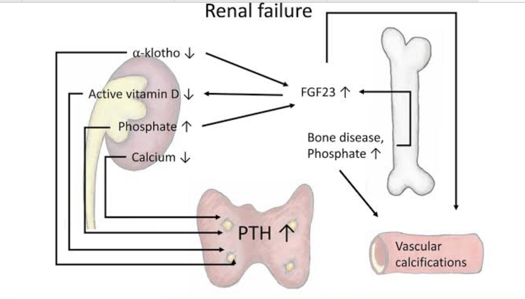
Chronic Kidney/Renal Failure Osteoporosis

16.

A 26-year-old woman was brought into the maternity department. Considering the term of her pregnancy (40 weeks), she should have gone into labor already. Examination determined that the uterine cervix is open but uterine contraction are absent. The doctor gave her a hormone drug for intensification a labor activity. Name this drug:


Explanation

Clearly, in this patient, there is inadequate propulsive power to cause fetal descent, cervical dilatation, and eventual expulsion of the fetus(es) and placenta.

Medical management: These are interventions that improve the quality of uterine contractions.

Oxytocin:

Provided there are no contraindications. Oxytocin is the medication of choice for augmenting contractions. The dosage regimen should be titrated to effect for achieving adequate uterine contractions. The usual protocol is 5units of oxytocin in 500mls of 5% Dextrose intravenous infusion, starting with 10 drops/min and gradually titrating the rate to achieve a contraction rate of at least 3 per minute. 

Amniotomy:


Membrane rupture (amniotomy) stimulates contractions by the release of prostaglandins and reflex stimulation of the uterus when the presenting part becomes closely applied to the lower uterine segment.

17.

A test animal received electrical impulses that irritate the sympathetic nerve that innervates blood vessels of the skin. What reaction will it cause in the blood vessels?

Explanation

The autonomic nervous system (ANS), comprised of two primary branches, sympathetic and parasympathetic nervous system, plays an essential role in the regulation of vascular wall contractility and tension. The focus is largely on the sympathetic nerves, which have a dominant role in cardiovascular control due to their effects to increase cardiac rate and contractility, cause constriction of arteries and veins, cause release of adrenal catecholamines, and activate the renin-angiotensin-aldosterone system.


Norepinephrine released from the sympathetic nerve terminals binds to α1- or α2-adrenergic receptors located on vascular smooth muscle cells to increase intracellular Ca2+. This rise in Ca2+ causes contraction of the smooth muscle.

Sympathetic effect Vasoconstriction

18.

A 50-year-old man complains of a hoarse voice and difficult breathing. Examination detected a laryngeal tumor in the region of the vocal cords. In this case metastases can spread to the following regional lymph nodes:


Explanation

Lymphatic drainage is important to understand due to the aggressive metastasis of some laryngeal cancers. The lymphatics of the larynx above the vocal cords drain initially to the infrahyoid nodes or directly to the deep cervical chain. The laryngeal lymphatic vessels below the vocal cords drain to the prelaryngeal and pretracheal nodes or directly to the deep cervical chain.

The deep (cervical) lymph nodes receive all of the lymph from the head and neck –  either directly or indirectly. They are numerous in number, but include the prelaryngeal, pretracheal, paratracheal, retropharyngeal, infrahyoid, jugulodigastric (tonsilar), jugulo-omohyoid and supraclavicular nodes.

The superficial cervical lymph nodes can be divided into the superficial anterior cervical nodes and the posterior lateral superficial cervical lymph nodes. The anterior nodes collect lymph from the superficial surfaces of the anterior neck. The posterior nodes collect lymph from superficial surfaces of the neck.

Submandibular nodes are located below the mandible in the submandibular triangle and collect lymph from the cheeks, the lateral aspects of the nose, upper lip, lateral parts of the lower lip, gums and the anterior tongue. They also receive lymph from the submental and facial lymph nodes.


Submental: collect lymph from the central lower lip, the floor of the mouth and the apex of the tongue.
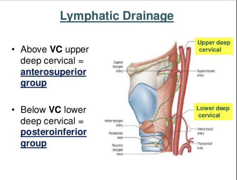
Vocal cords lymphatic drainage VC: Vocal Cord
19.

Clinical instrumental examination of a patient allowed to provisionally diagnose him with liver cancer. What protein is indicative of this diagnosis, if it is present in the blood serum?

Explanation

Once a suspicion of liver cancer arises, a physician will order one the following to confirm a diagnosis:

  1. Blood tests: alpha-fetoprotein (AFP), which may be elevated in 70% of patients with liver cancer. AFP levels could be normal in liver cancer. A rising level of AFP is suspicious for liver cancer. 

  2. Imaging studies: CT scan and MRI with contrast of the liver are the preferred imaging for detecting the location and extent of blood supply to the cancer. 

  3. Liver biopsy is performed to sample tissue from the lesion in the liver, which is analyzed by a pathologist to confirm the suspected diagnosis of liver cancer.

Gamma globulins: are Immunoglobulins or Antibodies.

Paraproteins: commonly found in Multiple Myeloma

Properdin: commonly found as part of the Complement system helping to clear immune complexes.


C-reactive protein: is an acute phase protein that can be elevated in Inflammatory conditions.

20.

10 days after the administration of an antitoxic anti-diphtheritic serum, a child with diphtheria developed skin rashes accompanied by severe itching, body temperature increased up to 38 degree C, the child developed pain in the joints. What likely cause of these phenomena can be suggested?

Explanation

Serum sickness is an immune-complex-mediated hypersensitivity reaction that classically presents with fever, rash, polyarthritis or polyarthralgias. Antibodies to foreign proteins are produced and 1 - 2 weeks later, antibody-antigen complexes form and deposit in tissues. The symptoms of serum sickness arise as a result of the formation of immune complexes between human proteins and heterologous (nonhuman) proteins. Medications containing heterologous antigens are the most common cause of serum sickness and include vaccinations (i.e., Rabies), immune modulating agents (i.e., rituximab, infliximab) and anti-venoms (antibodies against snake venom) or just as we have in this case - anti-diphtheric serum (antibodies against diphtheria toxin).

Serum sickness is a classic example of a Type III (Immune complex) Hypersensitivity reaction.

Anaphylactic and Atopy: Type I (Immediate) Hypersensitivity reaction.


Contact allergy and Delayed Type: Type IV Hypersensitivity reaction.

21.

Parents of a sick child came to the infectious disease specialist. The parents were working in India for a long time. The child has the following signs: a gray tint to the skin tone, loss of appetite, inertness, enlarged liver, spleen and lymph nodes. What protozoan disease can be suspected in the child?


Explanation

Leishmaniasis is due to protozoan parasites from the Leishmania species from being bitten by an infected sand fly. The sand flies that carry the parasite typically reside in tropical and subtropical environments. Fatal epidemics have occurred in areas of Asia, East Africa, and South America.

Leishmaniasis comes in three forms: cutaneous, visceral, and mucocutaneous.

Visceral leishmaniasis is sometimes known as systemic leishmaniasis or kala azar. It usually occurs two to eight months after being bitten by a sand fly. It damages internal organs, such as your spleen and liver. It also affects your bone marrow, as well as your immune system through damage to these organs.

Lambliasis is caused by Giardia lamblia, presents with diarrhea and foul smelling stool. 

Toxoplasmosis caused by Toxoplasma gondii, found in cat feces, presents with muscle pain, fever and headache.

Balantidiasis, caused by Balantidium coli, commonly found in pigs and in humans, it presents with gastrointestinal symptoms like chronic diarrhea, abdominal pain etc.


Amebiasis, caused by Entamoeba histolytica, presents with loose stool, abdominal cramping, and stomach pain.

22.

ECG of the patient shows increased duration of the QRS complex. What is the most likely causes?


Explanation

An electrocardiogram, or ECG, is a recording of the heart’s electrical activity as a graph over a period of time. A typical ECG tracing of the cardiac cycle (heartbeat) consists of a P wave (atrial depolarization), a QRS complex (ventricular depolarization), and a T wave (ventricular repolarization). 

The first wave on an ECG is the P wave, indicating atrial depolarization in which the atria contract (atrial systole). The QRS complex refers to the combination of the Q, R, and S waves, and indicates ventricular depolarization and contraction (ventricular systole).The T Wave indicates ventricular repolarization, in which the ventricles relax following depolarization and contraction. 

Therefore, an increased duration of the QRS complex corresponds to an Increased period of ventricular excitation.


The flat line (Isoelectric line) between the P wave and the beginning of the QRS complex indicates the conduction through the AV node.

Increased atrial and ventricular excitability will basically affect both the P wave and the QRS complex. (NB: the question only mentioned the QRS complex).


Increased period of atrial excitation = prolonged P wave
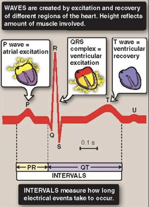
ECG Ventricular Excitation
23.

Blood test shows the following: Sodium – 115 mmol/L, chlorides – 85 mmol/L, glucose – 6 mmol/L, total protein – 65 g/L. The first consequence of such changes will be the decrease of:

Explanation

Check the table below for complete laboratory values - Biochemical Blood Analysis

Sodium - 115mmol/L (Norm: 136 - 145)

Chlorides - 85mmol/L (Norm: 95 - 105)

Glucose - 6mmol/L (Norm: 3.3 - 6.1)

Total Protein - 65g/L (Norm: 60 - 78)


From this, it is evident that there is a low Sodium and Chloride level in the blood of this patient. So, what is the consequence of a low Sodium chloride (NaCl)?

Sodium is the major cation of the extracellular fluid. It is responsible for one-half of the osmotic pressure gradient that exists between the interior of cells and their surrounding environment.

Chloride is also a predominant extracellular anion. Chloride is a major contributor to the osmotic pressure gradient between the Intracellular fluid (ICF) and Extracellular fluid (ECF), and plays an important role in maintaining proper hydration. 

Therefore, a decrease in both will result in a decrease in osmotic blood pressure.


Oncotic blood pressure is maintained by Albumin, Globulin and other plasma proteins.

Erythrocyte Sedimentation Rate is often increased in chronic inflammatory conditions.


Blood pH is controlled by acids and bases (goes up with increased bases and down with increased acids).

Laboratory Values 1 Laboratory Values 2
24.

A patient complains of frequent and excessive urination and thirst. Urinalysis revealed the following: 24 hour diuresis – 19 liters, specific gravity – 1.001. These values are characteristic of:


Explanation

Urine specific gravity: 1.001 (Norm: 1.016 - 1.022) 

This specific gravity is an indication of a very dilute urine. (NB: If it's low - Dilute; If it's high - concentrated urine).


Diabetes insipidus is a rare disorder that occurs when a person's kidneys pass an abnormally large volume of urine that is insipid—dilute and odorless. In most people, the kidneys pass about 1 to 2 litres of urine a day. In people with diabetes insipidus, the kidneys can pass 3 to 20 litres of urine a day. As a result, a person with diabetes insipidus may feel the need to drink large amounts of liquids.

Diabetes Insipidus is a disorder of the Vasopressin/Antidiuretic Hormone (ADH) regulation and function. It can be subdivided into Central and Nephrogenic.

Central - deficiency of ADH

Nephrogenic - ADH is present but the kidney does not respond.

Diabetes insipidus and diabetes mellitus—which includes both type 1 and type 2 diabetes—are unrelated, although both conditions cause frequent urination and constant thirst. Diabetes mellitus causes high blood glucose, or blood sugar, resulting from the body's inability to use blood glucose for energy. People with diabetes insipidus have normal blood glucose levels; however, their kidneys cannot balance fluid in the body.

25.

A 16-year-old girl has no hair on the pubis and in the armpits, her mammary glands are underdeveloped, no menstruation. This condition can be caused by the following hormone imbalance:


Explanation

The period between puberty and menopause represents the reproductive window of most women. If the ovaries stop working prematurely (roughly before the age of 40) and there are subsequently no menstrual periods, this is called premature ovarian failure. When this happens, your ovaries don't produce normal amounts of the hormone estrogen or release eggs regularly. This condition often leads to infertility. 

In the absence of ovarian hormones, secondary sexual characteristics like pubic hair growth and breast enlargement will be absent as well.

In females, breasts are a manifestation of higher levels of estrogen; estrogen also widens the pelvis and increases the amount of body fat in hips, thighs, buttocks, and breasts. Estrogen also induces growth of the uterus, proliferation of the endometrium, and menstruation. 


Estrogen that is responsible for all these, is produced in the Ovary. When there is ovarian failure, there will be decreased or absent estrogen which will result in the manifestations seen in this patient.

Pancreatic Islet failure - Diabetes Mellitus

Hyperthyroidism - Heat intolerance, weight loss, diarrhea, tachycardia

Hypothyroidism - Cold intolerance, weight gain, constipation, bradycardia

Adrenal medulla hyperfunction - a good example of this is Pheochromocytoma - hyperfunction of the sympathetic nervous system - hypertension, excessive sweating etc.

26.

A patient has an allergic response with itching, edemas and skin redness. In the tissues there is an increased concentration of a certain biogenic amine. Name this biogenic amine:

Explanation

Histamine, which is stored mainly in mast cells and basophils, is a prominent contributor to allergic disease. Elevations in plasma or tissue histamine levels have been noted during anaphylaxis and experimental allergic responses of the skin, nose, and airways. In the skin the cardinal features of urticaria (vasodilation - redness, vascular permeability - edema, and pruritus - itching) can be mediated by stimulation of the H1 receptor. H2 receptors are found in the stomach and it is not involved in allergic responses.
27.

It is known that hepatitis D virus belongs to defective viruses and can reproduce in the host cells only in the presence of another hepatitis virus. Name this other hepatitis virus:


Explanation

Hepatitis D virus (HDV) is a defective virus that depends on hepatitis B virus (HBV) to supply envelope proteins (HBsAg - Hepatitis B Surface Antigen) for assembling a new virion.


Four hepatitis viruses (HAV, HBV, HCV, and HEV) can infect humans independently, but HDV cannot. HDV either coinfects or superinfects with HBV because it requires the support of HBV to complete its life cycle. Therefore, HDV is known as a defective virus, or a satellite virus of HBV.

28.

Genealogical analysis of a child with myotonic dystrophy determined that this disease manifests in every generation, is in equal measure present in the relatives of both genders, the risk of inheriting this disease is equal no matter which parent is affected. If one of the parents is heterozygous for this disease and the other parent is healthy, the risk of them giving birth to a sick child is 50%. What type of disease inheritance is it?

Explanation

Key points to note from the question are - manifests in every generation; affects both genders equally; One parent is Heterozygous (Aa)


Autosomal Dominant:

  • Affects every generation - Grandparents, Parents, Children

  • Affects both male and female

  • With one affected (heterozygous) parent, on average, ½ or 50% of children will be affected.


Autosomal Recessive:

  • Skips generation - Grandparents are affected, Parents are healthy (skips parents), Children are affected

  •  Both parents are usually carriers (heterozygous), on average, ¼  or 25% of children will be affected; another 25% will not be affected and are not carriers; then 50% will be carriers


X-linked Dominant:

  • Transmitted through both parents

  • Mothers transmit to 50% of daughters and sons

  • Fathers transmit to all daughters but no sons


X-linked Recessive:

  • No male-to-male transmission i.e. Fathers can’t transmit to sons

  • Mother can transmit to 50% of sons


Y-linked:

  • Only possible in male patients XY

29.

A certain vitamin is a component of glutamic acid decarboxylase as a coenzyme, takes part in the formation of gamma-aminobutyric acid and its deficiency manifests as convulsions. Name this vitamin:


Explanation

Vitamin B6 also known as Pyridoxine - the active form is Pyridoxal phosphate.

Pyridoxal phosphate is a cofactor used in decarboxylation (e.g. Glutamate decarboxylase) and transamination reactions (e.g. AST and ALT). These reactions lead to the synthesis of Heme, Niacin, Histamine, Neurotransmitters like GABA, serotonin, dopamine, Epinephrine and Norepinephrine.

Glutamate decarboxylase (GAD) catalyzes the irreversible decarboxylation of L-glutamate to the valuable food supplement γ-aminobutyric acid (GABA). GAD requires pyridoxal phosphate (PLP) for its glutamate decarboxylase activity. GABA plays a major role as an inhibitory neurotransmitter in the central nervous system.


Deficiency of Vitamin B6 (Pyridoxine) can manifest as Convulsions, hyperirritability, peripheral neuropathy and anemia.

Amino Acid Derivatives

BH4 - Folic acid

B6 - Pyridoxine

30.

Histological specimen shows parenchyma of an organ that consist of lymphoid tissue that forms lymph nodules; the nodules are located diffusely and have a central artery. What anatomical structure has such morphological characteristic?


Explanation

Histologically, the spleen is comprised of red pulp and white pulp.  The red pulp is the site of blood filtration and the white pulp is lymphoid tissue that responds to blood-borne antigens.

Unlike the thymus and lymph nodes, the spleen lacks division into the cortex and medulla.   Lymphatic nodules with or without germinal centers (B cells) and with prominent eccentric central arterioles (called “central arteries) may be observed randomly distributed throughout the splenic pulp.  These nodules together with areas of dense lymphatic tissue surrounding central arterioles, called the periarterial lymphatic sheaths (PALS), comprise the splenic white pulp. This lymphatic sheath is made up of T cells.


Of all the answer choices listed, only the spleen has a central artery.

31.

A medical student was hospitalized into the infectious diseases unit on the 2nd day after the disease onset. The patient is suspected to have infectious mononucleosis. What results of laboratory analysis can confirm this diagnosis immediately on the day of the hospitalization.


Explanation

Infectious mononucleosis is caused by Epstein Barr Virus.

Testing for IgM-class antibodies is commonly performed to assist in the diagnosis of acute or reactivated infection. For a number of viral infections, including Epstein-Barr virus (EBV), measles and parvovirus B19, by the time patients present clinically, IgM levels are typically high, whereas viral loads have typically declined below detectable levels. Serologic testing, and specifically testing for IgM-class antibodies, is therefore a routine diagnostic approach for these viruses, particularly for immunocompetent individuals. 


IgG indicates a chronic infection; resolved infection or vaccination.

32.

After a spinal trauma the patient was lost proprioceptive sensitivity in the lower half of the body and in the lower limbs. What sensory conduction pathway was likely damaged, leading to the loss of sensitivity?

Explanation

The dorsal column of the spinal cord is formed by two large fasciculi (bundles of nerve fibers) running through the posterior spinal cord: fasciculus gracilis and fasciculus cuneatus. 

The gracilis fasciculus, also known as the “column of Goll”, carries tactile and proprioceptive information from the lower half of the body (T7 to the first coccygeal nerve). It lies medially to fasciculus cuneatus within the posterior half of the posterior spinal cord.

The fasciculus cuneatus, also known as the “column of Burdach”, represents the lateral portion of the dorsal column. It carries input from C1 and T6 spinal cord levels. This part of the Dorsal column of the spinal cord is responsible for transmitting vibration, conscious proprioception and fine (discriminative) touch sensations from the upper body.


The other pathways listed are not found in the posterior column.

Anterior and Lateral Spinothalamic tract: carries pain and temperature (not proprioception).


Spinocerebellar tract (Gowers): responsible for posture and coordination of movement.

Spinal Cord Pathways Spinocerebellar Gracilis Cuneatus
33.

A patient presents with an acute attack of cholelithiasis. Laboratory examination of the patient’s feces will show the following in this case:


Explanation

Bilirubin Jaundice Direct Indirect Stercobilin

We will be using this image to explain this question.

Red Blood Cells breakdown and release Heme from hemoglobin. Heme is broken down to unconjugated bilirubin. Unconjugated bilirubin goes to the liver and becomes conjugated. It leaves the liver and enters the intestine where it is converted to Stercobilin (it gives feces its color). A small percentage also enters the blood and is converted to urobilin (gives urine its color).

Cholelithiasis is the formation of gallstones which eventually blocks the cystic duct leading to mechanical/post-hepatic/obstruction/Direct jaundice.

Once there is a block, it prevents conjugated bilirubin from getting to the intestine. Therefore, Stercobilin cannot be made. Hence, we’ll have a negative reaction to Stercobilin.

If there is no blockage, we’ll have a positive reaction to Stercobilin.

34.

Autopsy of the body of 72-year-old woman, who was suffering from rheumatoid arthritis and died of uremia, revealed enlarged pale gray kidneys with glossy lardaceous surface on section. Based on the macroscopic changes, the pathologist suspected:

Explanation

Secondary or reactive amyloidosis occurs as a complication of chronic infectious or noninfectious inflammatory conditions associated with tissues destruction such as Tuberculosis, Autoimmune disorders (Rheumatoid arthritis, dermatomyositis, scleroderma), Chronic Osteomyelitis, Inflammatory bowel disease (Ulcerative colitis and Crohn’s disease) etc.

Secondary amyloidosis is typically distributed in solid abdominal organs like the liver, kidney, spleen and adrenals. In renal amyloidosis, the kidneys may be normal-sized, enlarged or contracted. Cut surface is pale waxy and translucent.


From the information given in the question stem: 72 year old woman suffering from Rheumatoid arthritis (a predisposing condition for secondary amyloidosis) with an enlarged, glossy lardaceous kidney, it is safe to suspect a Renal Amyloidosis here.

35.

The patient notes frequent diarrheas, especially after eating fatty food and loss of weight. Laboratory testing detected steatorrhea and hypocholic feces. What is the likely cause of this condition?


Explanation

Red Blood Cells breakdown and release Heme from hemoglobin. Heme is broken down to unconjugated bilirubin. Unconjugated bilirubin goes to the liver and becomes conjugated. It leaves the liver and enters the intestine where it is converted to Stercobilin (it gives feces its color). A small percentage also enters the blood and is converted to urobilin (gives urine its color).

The conjugated Bilirubin that goes to the intestine is needed for fat digestion. 

In the case of a formation of gallstones, cancer of the pancreatic head or any other condition that blocks the cystic duct or common bile duct, this will result in mechanical/post-hepatic/obstruction/Direct jaundice.


Once there is a block, it prevents conjugated bilirubin from getting into the intestine. Stercobilin cannot be made (i.e. feces will be pale/acholic or hypocholic) and there will also be fat indigestion (steatorrhea).

Bilirubin Jaundice Direct Indirect Stercobilin

36.

A person developed increased pulmonary ventilation due to physical exertion. What indicator of external respiration will be significantly increased compared to the resting state?


Explanation


Lung volumes are also known as respiratory volumes. It refers to the volume of gas in the lungs at a given time during the respiratory cycle. So if there is an increased lung ventilation, it therefore implies that compared to the resting state, the respiratory volumes will also increase.

37.

A 48-year-old man is unconscious. He has a history of several syncopal episodes with convusions. ECG shows deformed QRS complexes unconnected with P waves, atrial contractions are approximately 70/min, ventricular contractions are 25-30/min. Name the type of arrhythmia in this case:

Explanation

Key point here is: we have a deformed QRS complexes UNCONNECTED with P waves.

This is the only information you need to answer this question.

Now let’s compare the answer choices. I won’t bore you with ECG images to avoid confusions.


Atrioventricular (AV) block is partial or complete interruption of impulse transmission from the atria to the ventricles. We have 3 degrees of heart block.

First Degree AV block: All normal P waves are followed by QRS complexes, but the PR interval is longer than normal (>0.20 second).

Second Degree AV block: Some normal P waves are followed by QRS complexes, but some are not. Two types exist:

  • Mobitz type I

  • Mobitz type II

In Mobitz type I 2nd-degree AV block, the PR interval progressively lengthens with each beat until the atrial impulse is not conducted and the QRS complex is dropped (Wenckebach phenomenon); AV nodal conduction resumes with the next beat, and the sequence is repeated.

In Mobitz type II 2nd-degree AV block, the PR interval remains constant. Beats are intermittently nonconducted and QRS complexes dropped, usually in a repeating cycle of every 3rd (3:1 block) or 4th (4:1 block) P wave.

Third Degree AV block: Heart block is complete in 3rd-degree AV block. There is no relationship between P waves and QRS complexes (AV dissociation) in 3rd-degree AV block. The P wave rate is greater than the QRS rate. (P waves 70/min; QRS-ventricular contractions 25-30/min).

There is no relationship between P wave and QRS complexes also means P wave is UNCONNECTED with QRS complexes.

 
38.

An oncology patient is to undergo a surgery on the descending colon. Name the main source of the blood supply to this organ:

Explanation

The superior mesenteric artery (SMA) and the inferior mesenteric artery (IMA) provide blood supply to the colon.

The cecum, ascending colon and transverse colon (middle colic, which is a branch of Superior Mesenteric artery) receive their blood supply from the superior mesenteric artery.

The descending and sigmoid colon receive their blood supply from the left colic and sigmoid arteries, which are branches of the Inferior Mesenteric Artery.

Celiac trunk supplies the stomach (Left gastric artery), liver (common hepatic artery) and spleen (splenic artery).

So, the main blood supply to the descending colon is the Inferior mesenteric artery.

39.

Body fluids, especially urine, of a sick child produce a specific sweet odor. It is associated with the disturbed metabolism of such amino acids as leucine, isoleucine and valine. What diagnosis will be made by the doctor in this case?

Explanation

Elevated concentrations of branched-chain amino acids (leucine, isoleucine, and valine), as well as a generalized disturbance of amino acid concentration ratios, are present in blood and the maple syrup odor can be detected.

Maple syrup urine disease (MSUD) is caused by decreased activity of the branched-chain alpha-ketoacid dehydrogenase complex (BCKD), the second enzymatic step in the degradative pathway of the branched-chain amino acids (BCAAs), which includes leucine, isoleucine, and valine.

Alkaptonuria (ochronosis): accumulation of Homogentisic acid due to deficiency of Homogentisate dioxygenase. Patients have dark urine or urine that turns black when exposed to air.

Phenylketonuria: accumulation of Phenylalanine due to deficiency of Phenylalanine hydroxylase. It can lead to intellectual disability, seizures, behavioral problems, and mental disorders with a musty smell and lighter skin.

Fructosuria: deficiency of Fructokinase or Aldolase B, accumulation of Fructose or Fructose-1-phosphate.

Galactosemia: deficiency of Galactokinase, accumulation of Galactose.

40.

Examination of the femoral bone detected chronic suppurative inflammation of the compact substance and bone marrow, formation of bone sequestra. What disease is associated with such changes?


Explanation

Chronic osteomyelitis represents a progressive inflammatory process caused by pathogens, resulting in bone destruction and sequestrum formation. Late signs include increased bone resorption, formation of sequestra and new bone formation in the periosteum or endosteum.


A sequestrum (plural: sequestra) is a piece of dead bone that has become separated during the process of necrosis from normal or sound bone. It is a complication (sequela) of osteomyelitis.

41.

Autopsy of the body revealed a large wedge-shaped patch of dense dark red tissue with clear margins in the upper lobe of the right lung. Histological examination detected there necrosis of the alveolar walls; the alveolar lumen is tightly packed with erythrocytes. What process occurred in the lungs?

Explanation


Pulmonary infarction (PI) is usually a complication of another primary disease state, most commonly a pulmonary embolism (PE). An x-ray image may be the first clue toward a diagnosis of pulmonary infarction. Hampton’s hump (wedge-shaped consolidation at the lung periphery), Westermark’s sign (radiographic oligemia or increased lucency), and Fleischer sign (prominent pulmonary artery) are specific findings. The red infarct is wedge-shaped and based on the pleura. These infarcts are hemorrhagic because, though the pulmonary artery carrying most of the blood and oxygen is cut off, the bronchial arteries from the systemic circulation (supplying about 1% of the blood to the lungs) is not cut off.

42.

A nurse mistakenly gave nearly a double dose of insulin to a patient with diabetes mellitus, which caused hypoglycemic coma in the patient. What medicine should be administered to bring the patient out of the coma?


Explanation

For a patient in hypoglycemic coma, essentially, a quick-acting carbohydrate needs to be given, followed by a longer-acting carbohydrate. Initially, Glucose 10-20 g is given by mouth, either in liquid form or as granulated sugar (two teaspoons) or sugar lumps.

Repeat capillary blood glucose after 10-15 minutes; if the patient is still hypoglycaemic then the above can be repeated (probably up to 1-3 times).


If the patient is unconscious or uncooperative, Intravenous administration of 75-80 ml 20% glucose or 150-160 ml of 10% glucose. Also, Glucagon 1 mg should be given by intramuscular (IM), or subcutaneous (SC) injection can be administered.

43.

A lab rat has subcutaneously received mercury(II) chloride in the amount of 5 mg/kg. 24 hours later the plasma creatinine concentration increased several times. What mechanism of retention azotemia is observed in this case?


Explanation


The kidneys are sensitive to the effects of mercury, because mercury accumulates in the kidneys and causes higher exposures to these tissues, and thus more damage. All forms of mercury can cause kidney damage if large enough amounts enter the body. Mercury related poisoning is implicated in  nephrotoxic Acute Renal Failure. And Acute kidney injury (AKI), formerly called acute kidney failure, is a sudden decline in glomerular filtration rate (GFR).

44.

In the hospital a patient was diagnosed with anemia. During the anamnesis collection, it was revealed that the patient had been suffering from peptic ulcer disease of the stomach for several years already. What type of anemia is most likely in this patient?


Explanation

Peptic ulcers occur when acid in the digestive tract eats away at the inner surface of the stomach or small intestine. The acid can create a painful open sore that may bleed. If left untreated, peptic ulcers can result in Internal bleeding. Bleeding can occur as slow blood loss that leads to anemia or as severe blood loss that may require hospitalization or a blood transfusion. Severe blood loss may cause black or bloody vomit or black or bloody stools. Chronic posthemorrhagic anemia occurs with prolonged moderate hemorrhage as seen in this patient with a history of peptic ulcer disease for several years.


Acute posthemorrhagic anemia will be inappropriate in this case because of the history spanning several years already.

45.

24 hours after an appendectomy the patient’s blood test shows neutrophilic leukocytosis with a regenerative shift. What is the most likely mechanism of absolute leukocytosis development in the patient’s peripheral blood?


Explanation

Neutrophilic leukocytosis with a regenerative shift also means neutrophilia with left shift. Neutrophilia refers to a higher than normal number of neutrophils. Left shift describes when immature neutrophils are released from the bone marrow. In any acute inflammation (e.g. acute appendicitis), an increase in neutrophils is often seen. Increases may also be seen after a heart attack (or other infarct) and necrosis.

Leukopoiesis is the production of new White Blood Cells and this process takes place in the bone marrow. The normal reaction of bone marrow to infection or inflammation leads to an increase in the number of white blood cells, predominantly polymorphonuclear leukocytes (neutrophils) and less mature cell forms (the regenerative shift or “left shift”).

In this case, intensification of leukopoiesis led to the Neutrophilic leukocytosis with a regenerative shift. Absolute leukocytosis as mentioned in the question, also points to increased (intensification) of Leukopoiesis.


The other options listed: Leukocyte redistribution, Deceleration of leukocyte migration to the tissues, decreased leukocyte disintegration and immunity activation are all a product of relative Leukopoiesis (absolute leukopoiesis has to do with production of new blood cells - Leukopoiesis).

46.

A patient with massive burns received a skin graft from a donor. On the 8th day after the grafting, the transplant became edematous and discolored. On the 11th day transplant rejection started. What cells take part in this process?

Explanation

Transplant rejection can be: Hyperacute; Acute or Chronic.

Hyperacute: occurs within minutes, most times while still in the operating room. It is mediated by pre-existing antibodies (B cells) in the recipient - Type II hypersensitivity reaction.

Acute: occurs within weeks to months. It is mediated by T cells - Type IV hypersensitivity reaction.

Chronic: occurs in months to years. It is mediated by T cells (mainly CD4+ T cells). Both cellular and humoral components are involved here - Type II and IV hypersensitivity reaction.

In this patient, the graft rejection became manifest on the 8th day (1 week) which meets the criteria for an acute transplant rejection mediated by T cells.

47.

A 15-year-old teenager complains of general weakness, dizziness and rapid fatigability. Examination detected deformed erythrocytes, their number is decreased. The provisional diagnosis of sickle-cell anemia was made. What amino acid replacement in hemoglobin causes the development of this pathological condition?

Explanation

Sickle cell anemia results from the single amino acid substitution of valine for glutamic acid in the beta-chain owing to a nucleotide defect that causes the production of abnormal beta-chains in hemoglobin S. Abnormal hemoglobin chains form polymers in the deoxygenated state, leading to the characteristic sickle cells. The polymerization of deoxygenated hemoglobin S accounts for the pathologic changes in sickle cell disease.

What this means is that: Glutamate is taken out and Valine is used instead (Glutamate replaced with Valine).


Not to be confused with the other option that says - Valine replaced with Glutamate.

48.

Name the supramolecular multienzyme complex that is integrated into the lipid layer of inner mitochondrial membrane that creates conditions for redox reactions:

Explanation

Mitochondria are the powerhouses of the cell. Biological energy conversion in mitochondria is carried out by the membrane protein complexes of the respiratory chain and the mitochondrial ATP synthase in the inner membrane cristae.

Mitochondria are separated from the cytoplasm by the outer and inner mitochondrial membrane. The outer membrane is porous and freely traversed by ions and small, uncharged molecules. The inner membrane is where oxidative phosphorylation takes place in a suite of membrane protein complexes of the respiratory chain that create the electrochemical gradient across the inner membrane, or use it for ATP synthesis.

49.

The process of tissue respiration is accompanied by oxidation of organic compounds and synthesis of macroergic molecules. In what organelles does this process occur?

Explanation

Oxidation of organic compounds and synthesis of macroergic molecules simply means oxidative phosphorylation and synthesis of ATP - these processes take place in the Mitochondria.


Mitochondria are separated from the cytoplasm by the outer and inner mitochondrial membrane. The outer membrane is porous and freely traversed by ions and small, uncharged molecules. The inner membrane is where oxidative phosphorylation takes place in a suite of membrane protein complexes of the respiratory chain that create the electrochemical gradient across the inner membrane, or use it for ATP synthesis.

Lysosomes are responsible for digestion/break down substances; Ribosomes are responsible for protein synthesis; Golgi body or apparatus is responsible for packaging proteins for export out of the cell; Peroxisomes are responsible for Beta oxidation of fatty acids and detoxification.

50.

A patient with ischemic heart disease has increased levels of triglycerides and low density lipoproteins in this blood plasma. What medicine should he be prescribed?

Explanation

Fibrates are a class of drugs utilized in treating and managing serum cholesterol levels. Examples of fibrates: Gemfibrozil; Fenofibrate; Clofibrate etc. They help improve cholesterol by lowering triglycerides and raising HDL cholesterol.

There are two main types of cholesterol: high-density lipoprotein (HDL) and low-density lipoprotein (LDL). HDL cholesterol is considered the “good” type of cholesterol. LDL, on the other hand, is considered the “bad” type of cholesterol.

The only lipid lowering drug out of all the answer choices given is Fenofibrate.


Lisinopril: Angiotensin Converting Enzyme Inhibitor (ACE Inhibitor) Used in hypertension.

Amiodarone: an Antiarrhythmic drug (Class III - potassium blocker). It has no effect on cholesterol.

Famotidine: H2 histamine receptor blocker, useful in reducing gastric acid secretion to treat peptic ulcer disease and gastritis.

Dobutamine: Beta 1 receptor agonist used to improve cardiac contractility. It has no effect on lowering cholesterol levels.

 
51.

A woman with enteritis accompanied by severe diarrhea presents with the loss of water in the extracellular space, increased water content in the cells and decreasing blood osmolarity. Name this type of water-electrolyte imbalance:

Explanation

Key points to note:

Loss of water in the extracellular space: Hypohydration

Decrease blood osmolarity: Hypoosmolar

(NB - We are mainly concerned about the extracellular fluid because that is what we can measure).

The characteristic feature of all kinds of hypohydration is the negative fluid balance: the predominance of water loss over its intake by the organism. According to the osmolality of extracellular fluid three types of hypohydration are singled out: hypoosmolar, hyperosmolar and iso osmolar.

In hypoosmolar hypohydration the organism’s salt losses are predominant as compared to water losses and the decrease in extracellular fluid osmolality. Causes: hypoaldosteronism, continuous profuse sweating, recurrent or uncontrollable vomiting, profuse diarrhea just as the patient in question (for example, in enteritis, cholera or malabsorption syndrome) associated with the loss of intestinal juice containing K+, Na+, Ca2+ and other cations (loss of water - hypohydration; loss of ions - hypoosmolar).

52.

Autopsy of the body of a person, who died after an abdominal surgery, revealed numerous thrombi in the veins of the lesser pelvis. Clinically, thromboembolism syndrome was registered. Where should the pathologist search for thromboembolis?

Explanation

This question is here to test your anatomy knowledge.

Put this at the back of your mind:

  • Veins take blood to the heart

  • Arteries take blood away from the heart.

  • When there is an embolus, it lodges and blocks the first capillary bed it encounters (i.e. it will lodge in the first organ it encounters once it leaves the heart).


Let’s dissect this question together.

There is a thrombi in the lesser pelvis. Now, you know that all veins in the lower half of the body drain to the Inferior Vena Cava (IVC) and the veins of the upper half of the body drain into the Superior Vena Cava (SVC). Then the SVC and IVC both drain into the right side of the heart from where the blood goes to the lungs to be oxygenated.

If you go back to the points I told you to put at the back of your mind earlier.

From the third point, you can now see that the first organ to be encountered by the thromboemboli once it leaves the heart is the Lungs.


So the pathologist should search for the thromboemboli in the Pulmonary arteries.

Deep Vein Thrombosis Pulmonary Embolism
53.

A patient with essential hypertension developed a cough during the systematic treatment with a hypotensive drug. What drug of those listed below can cause such a side effect?

Explanation

Angiotensin-converting enzyme inhibitors (ACE-I) like Lisinopril, Enalapril etc., are widely used in diseases, such as hypertension, congestive heart failure, and myocardial infarction. A dry, tickly and often bothersome cough is the most common adverse effect of ACE inhibitors.

In normal physiology, angiotensin I is converted to active angiotensin II by ACE produced by the capillaries in the lungs. Angiotensin II has many physiological effects, such as increasing the resistance of blood vessels, causing adrenal cortex aldosterone release, and stimulating vasopressin which all lead to an increased blood pressure. So, by inhibiting the ACE, we are able to bring down the blood pressure.

However, angiotensin converting enzyme (ACE) is also involved in the breakdown of bradykinin. Once ACE inhibitors are used, bradykinin accumulates. The increased bradykinin irritates the airways and produces dry cough.

Lisinopril is the only ACEI listed as an answer choice.

Verapamil: Calcium channel blocker

Prazosin: alpha 1 receptor blocker

Hydralazine: vasodilator

Dibazol: vasodilator with analgesic effect
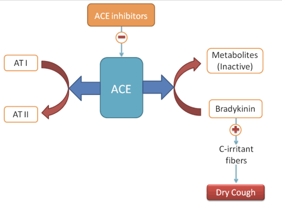
ACE Inhibitors Lisinopril Bradykinin Cough

54.

Pterin derivatives – aminopterin and methotrexate – are competitive inhibitors of dihydrofolate reductase. As a result, they suppress the regeneration of tetrahydrofolic acid from dihydrofolate. These medicines lead to the inhibition of intermolecular transport of one-carbon groups. In the process, the biosynthesis of the following polymer is suppressed:

Explanation


Dihydrofolate reductase is an enzyme that converts dihydrofolate to tetrahydrofolate and is involved in purines and thymidylate synthesis. Antifolate drugs, like methotrexate, can tightly bind to Dihydrofolate reductase and inhibit DNA, RNA synthesis and cell proliferation, eventually leading to cell death.

Inhibition of this pathway has a minimal effect on protein as compared to DNA and RNA synthesis because Methionine could be gotten from diet (e.g. there is high methionine, in egg, meat, fish etc.)

Methotrexate Folate antagonists Dihydrofolate reductase DNA synthesis

55.

A 2 year old child presents with marked delay in psychomotor development, vision and hearing deterioration, marked enlargement of the liver and spleen. The child is diagnosed with hereditary Neimann-Pick disease. What genetic defect is the cause of this disease?

Explanation

Niemann-Pick disease (NPD) is a lysosomal storage disease caused by acid sphingomyelinase deficiency, which catalyzes the hydrolysis of sphingomyelin to ceramide and phosphocholine. As a result, sphingomyelin and its precursor lipids begin to accumulate in lysosomes, mainly in macrophages. These lipid-laden macrophages deposit in the liver, spleen, lungs, and brain causing hepatosplenomegaly, cytopenias, lung disease, and neurologic symptoms.
56.

The patient presents with smoothed out nasolabial fold, dilated right palpebral fissures (It cannot be closed when squinting, because of eyelids would not close), there are difficulties during speaking and eating (Food gets stuck between the cheek and teeth). What nerve is damaged in this case?

Explanation

The facial nerve is a mixed nerve, it contains both sensory and motor fibers.

Take a look at the image below the explanation and Let’s pick each of the symptoms listed in the question one after the other.

  • Smoothed out nasolabial folds: facial nerve supplies most of the facial muscles and if those muscles fail to contract, there won’t be any fold (forehead, nasolabial folds). You will see that the right side on the image below is smoothed out.

  • Eyelids won't close: facial nerve supplies orbicularis oculi needed to close the eyelids. A damage to the facial nerve means the eyelid on that side of the face will remain open.

  • Food gets stuck between the cheek and teeth: facial nerve supplies the buccinator muscle of the cheeks, needed to push to the middle of the mouth so the tongue and roll it and swallow.


Trigeminal nerve supplies the four masticatory muscles needed to close and open the mouth which is not affected here. Abducens nerve (CN VI) supplies the lateral rectus muscle of the eyeball. All other nerves listed do not supply any muscle on the face.

Facial nerve bell's palsy krushkrok

57.

A woman was hospitalized in a severe condition with the following diagnosis; hemorrhagic stroke in the region of the medial surface of the right frontal lobe. What artery is likely to be damaged, causing this condition in the patient?

Explanation

Three main cerebral arteries supply the cerebral hemispheres.

Anterior cerebral artery supplies the medial surface of the frontal lobe and part of the parietal lobe.

Middle cerebral artery supplies the lateral surface of the parietal lobe and part of the lateral surface of the frontal lobe. It also gives branches to the internal capsule (deeper grey matter structures of the brain).

Posterior cerebral artery supplies the occipital lobe and the inferior surface of the temporal lobe.

Anterior communicating artery connects the right and left anterior cerebral arteries.


Posterior communicating arteries is a branch of the internal carotid artery. It connects the middle cerebral artery to the Posterior cerebral artery making the Circle of Willis a closed circuit.

Blood supply to the brain anterior cerebral artery middle cerebral artery

58.

In an experiment, a frog neuromuscular preparation had been processed with a curare-like substance, which led to the disappearance of muscle contractions in response to electrical stimulation. What function of the muscle cell membrane is disrupted by curare-like substance?


Explanation

Curare is the historical prototype of nondepolarizing neuromuscular blockers, but it is no longer used clinically. Curare was the first paralytic used in anesthesia, but it has been replaced by newer agents. 

They function in the neuromuscular synapse by competitively and reversibly inhibiting the nicotinic acetylcholine receptor, which is a subtype of acetylcholine receptor found at the neuromuscular junction. This causes weakness of the skeletal muscles and, when administered in a sufficient dose, eventual death by asphyxiation due to paralysis of the diaphragm.


This can be summarized as a receptor blocker, blocking the effect of the mediators (acetylcholine neurotransmitter) in the neuromuscular synapse. They only block the reception of the mediators in the neuromuscular synapse.

59.

An unconscious patient was brought into the admission room. He presents with cold skin, constricted pupils, difficult respiration with Cheyne-Stokes pattern, low blood pressure, overfilled urinary bladder. He was diagnosed with morphine poisoning. What drug should the patient be given as an antagonist in this case?

Explanation

Naloxone is an opioid antagonist medication used to block or reverse the effects of opioid drugs, particularly within the setting of drug overdoses which are rapidly becoming a leading cause of death worldwide. More specifically, naloxone has a high affinity for μ-opioid receptors, where it acts as an inverse agonist, causing the rapid removal of any other drugs bound to these receptors. When taken in large quantities, opioid medications such as morphine, hydromorphone, methadone, heroin, or fentanyl are capable of causing life-threatening symptoms such as respiratory depression, reduced heart rate, slurred speech, drowsiness, and constricted pupils.

Bemegride is a central nervous system stimulant.

Unithiol is used in poisoning with mercury and mercury salts.

Sodium thiosulfate is used to treat cyanide poisoning.


Cytitone has been used as a smoking cessation treatment.

Naloxone opioid antagonist morphine
60.

A patient has developed status epilepticus. What medicine should be used in this case to stop the seizures?

Explanation

Convulsive status epilepticus is a neurological emergency in adults and children. The benzodiazepines are some of the most effective drugs in the treatment of acute seizures and status epilepticus. The benzodiazepines most commonly used to treat status epilepticus are diazepam (Valium), lorazepam (Ativan), and midazolam (Versed). All three compounds work by enhancing the inhibition of γ-aminobutyric acid (GABA) by binding to the benzodiazepine-GABA and barbiturate-receptor complex.


Sodium bromide can be used as a hypnotic, as an anticonvulsant, and as a sedative. Even though Sodium bromide can be used as an anticonvulsant, drugs in the class of Benzodiazepines (diazepam, Lorazepam) have higher efficacy and potency.

Trihexyphenidyl is used to treat symptoms of Parkinson's disease or involuntary movements due to the side effects of certain psychiatric drugs.

Promethazine is used to treat anaphylaxis (sudden, severe allergic reactions) and the symptoms of the common cold such as sneezing, cough, and runny nose. Also to treat nausea and vomiting related to certain conditions (such as before/after surgery, motion sickness).

Valerian is most commonly used for sleep disorders, especially the inability to sleep (insomnia).

 
61. In the process of acetylcholine interaction with muscarinic acetylcholine receptors of contractile cardiomyocytes, a bioactive substance is being produced. This bioactive substance leads to inactivation of calcium channels, decrease of calcium ions entry into the cardiomyocytes and development of negative inotropic effect. Name this substance:

Explanation

Let’s take a careful look at the image below.

Sympathetic Nervous system which uses Norepinephrine (NE) and Epinephrine (Epi) as a neurotransmitter works to increase the heart rate.

Parasympathetic nervous system which uses Acetylcholine (ACh) as a neurotransmitter works to decrease the heart rate.


These two systems antagonize each other, while Norepinephrine works by activating an enzyme called Adenylyl cyclase (AC) which converts ATP to cAMP; Acetylcholine on the other hand inhibits this enzyme - Adenylyl cyclase thereby reducing the production of cAMP from ATP. And because cAMP is reduced, it decreases the influx of Ca2+ into cardiomyocytes thereby reducing cardiac contraction (decreased Inotropy).

Acetylcholine cAMP Norepinephrine Cardiomyocytes

62.

Examination showed that total leukocyte count in the patient’s blood is 11 x 109/L, with 80% neutrophils, among which 9% are band neutrophils. Characterize the changes in the cell composition of ‘white’ blood in this case:

Explanation

Neutrophilic leukocytosis with a regenerative shift also means neutrophilia with left shift. Neutrophilia refers to a higher than normal number of neutrophils (norm: 47 - 72%). Left shift describes when immature neutrophils are released from the bone marrow. In any acute inflammation (e.g. acute appendicitis), an increase in neutrophils is often seen. Increases may also be seen after a heart attack (or other infarct) and necrosis.

Leukopoiesis is the production of new White Blood Cells and this process takes place in the bone marrow. The normal reaction of bone marrow to infection or inflammation leads to an increase in the number of white blood cells, predominantly polymorphonuclear leukocytes (neutrophils) and less mature cell forms (the regenerative shift or “left shift”).


Immature neutrophils are called “bands”, and others use the term: “immature granulocytes” (norm: 1 - 6%)

Mature Neutrophils are called Segmented (Norm: 47 - 72%)

With a value of 80% for segmented and 9% for band neutrophils, then we definitely have a left shift indicated by the increased number of neutrophils.

63. Blood smear analysis of a blood sample obtained from a patient with inflammatory process shows a large number of round cells with segmented nucleus (three or more segments) and fine pink-violet granulation in the cytoplasm. Name the blood cells:

Explanation

Mature Neutrophils are called Segmented. They have 3 - 5 segments.

(NB: hypersegmented Neutrophils 6+ segments are seen in Vitamin B12 and folate deficiency).

In any acute inflammation (e.g. acute appendicitis), an increase in neutrophils is often seen. Increases may also be seen after a heart attack (or other infarct) and necrosis.

Leukopoiesis is the production of new White Blood Cells and this process takes place in the bone marrow. The normal reaction of bone marrow to infection or inflammation leads to an increase in the number of white blood cells, predominantly polymorphonuclear leukocytes (neutrophils) and less mature cell forms (the regenerative shift or “left shift”).


Erythrocytes (red blood cells): lack a nucleus and organelles. They are biconcave.

Basophils: contain densely basophilic granules (blue in color) - heparin and histamine.

Eosinophils: have a bilobed nucleus. Contain granules, pink in color.

Lymphocytes: B and T cells; round, densely staining nucleus with small amount of pale cytoplasm.

64. A newborn boy has dolichocephalic skull, microcephaly and defects of heart, kidneys and digestive system. The child’s karyotype is 47, XY + 18. Make the diagnosis:

Explanation

Edwards syndrome: Election age (18) - trisomy 18

Prominent occiput (dolichocephalic skull); rocker-bottom feet; intellectual disability; Microcephaly; congenital heart disease, omphalocele (digestive system).

Down syndrome: Drinking age (21) - Trisomy 21

Intellectual disability; single palmar crease; epicanthal folds; duodenal atresia; congenital heart disease

Patau syndrome: Puberty (13) - Trisomy 13

Severe intellectual disability; microcephaly; cleft lip and palate; polydactyly

Turner syndrome: Female, 45, XO, No Barr body

Short stature; ovarian dysgenesis (streak ovary); webbed neck

Klinefelter syndrome: Male, 47, XXY, 1 Barr body.

Testicular atrophy; tall; long extremities; gynecomastia;

 
65. A histological specimen demonstrates a vessel with the wall that consists of endothelium, basement membrane and loose connective tissue. What type of vessel is it?

Explanation

The veins have the three basic layers (tunica adventitia, tunica media and tunica intima).

Tunica Intima: A thin endothelial lining, (in some veins, you may be able to see the valves).

Tunica Media: This layer contains 2-3 layers of muscle cells.

Tunica Adventitia: This is the broadest layer. It contains longitudinal collagen fibres, and vasa vasorum.

From the question stem, the specimen being described consists of an endothelium and basement membrane (Tunica Intima); Loose connective tissue (Tunica adventitia).

You can see that the Tunica Media which has muscle cells is absent in this specimen which makes it a NON-MUSCULAR VEIN.

66. Arterial blood pH is 7.4; Primary urine pH is 7.4; final urine pH is 5.8. Decreased pH of the final urine results from the secretion of a certain substance in the nephron tubules. Name the substance:

Explanation

Normal pH can be said to be between 7.35 - 7.45.

Anything below this range is termed acidic (Acidosis) and above this range is termed Basic (Alkalosis).

So, a urine pH of 5.8 is clearly acidic and this is a result of increased acidic content in urine. One of the main contributing factors to this is the secretion of H+ in renal tubules.


The kidneys maintain normal acid-base balance primarily through the reabsorption of sodium and the tubular secretion of hydrogen and ammonium ions. Urine becomes increasingly acidic as the amount of Hydrogen ion and excess acid retained by the body increases.

67. A patient came to the hospital complaining of abdominal distension, diarrhea and meteorism that occur after eating protein-rich food, which indicated disturbed protein metabolism and intensified protein putrefaction. What substance is the product of this process in the intestine?

Explanation


Protein escaping the small intestine is subject to putrefaction in the caecum (large intestine). Putrefaction inside the human gastrointestinal tract (gut) pertains to decomposition or fermentation of undigested proteins by the resident microbiota. Once such undigested proteins are broken down into amino acids in the large intestine, they are usually metabolized by the resident proteolytic bacteria, leading to production of harmful metabolites. Such products include amines, indoles and ammonia.

68. A patient complains of acute increase in diuresis (upto 5-7 liters of urine per 24 hours). Examination of vasopressin in this patient. What cells have insufficient secretory activity in this case?

Explanation

Diabetes insipidus is a rare disorder that occurs when a person\'s kidneys pass an abnormally large volume of urine that is insipid—dilute and odorless. In most people, the kidneys pass about 1 to 2 litres of urine a day. In people with diabetes insipidus, the kidneys can pass 3 to 20 litres of urine a day. As a result, a person with diabetes insipidus may feel the need to drink large amounts of liquids.

Diabetes Insipidus is a disorder of the Vasopressin/Antidiuretic Hormone (ADH) regulation and function. It can be subdivided into Central and Nephrogenic.

Central - deficiency of ADH

Nephrogenic - ADH is present but the kidney does not respond.


From the question stem, it can be confirmed that there is decreased Vasopressin (ADH) which is clearly a case of Central Diabetes Insipidus. Now, the question is, where is ADH produced? Which cells are producing less than normal ADH?

ADH is produced by the hypothalamus in the brain and stored in the posterior pituitary gland at the base of the brain. Specialized neuron clusters called neurosecretory cells in the hypothalamus produce the hormones Antidiuretic Hormone (ADH) and Oxytocin (OXT), and transport them to the pituitary, where they\'re stored for later release.

69. A 35-year-old man complains of a severe rhinitis and a loss of sense of smell for a week. Examination detected in the nasal cavity a large amount of mucus that covered the mucosa and blocked the olfactory receptors. Where in the nasal cavity are these receptors located?

Explanation

The olfactory epithelium occupies about 1 to 2 cm2 of the roof of the nasal cavity. It covers the superior nasal concha, and lies directly beneath the cribriform plate of the ethmoid bone.

The olfactory nerves (Cranial Nerve I - CN I) are special sensory nerves for the sense of smell. They originate in the receptors of the olfactory epithelium and pass through the olfactory foramina in the cribriform plate of the ethmoid bone, ending at the olfactory bulbs. Specialized neurons called olfactory receptors are located in the epithelia that cover the roof of the nasal cavity, superior nasal conchae, and superior nasal septum.


The three lines of the Nasal conchae correspond to the Superior, Middle and Inferior Nasal Conchae respectively.

Olfactory epithelium Superior Nasal Concha

70.

A 50-year-old man experienced a severe stress. His blood levels of adrenaline and noradrenaline sharply increased. What enzyme catalyze the process of noradrenaline inactivation?

Explanation

Noradrenaline and Adrenaline are also known as Norepinephrine and Epinephrine respectively.

Monoamine oxidase (MAO) is an enzyme that degrades or breaks down three neurotransmitters associated with mood and anxiety. Monoamine oxidase breaks down norepinephrine, serotonin, and dopamine. These 3 compounds are called monoamines. 

Monoamine oxidases (MAOs) A and B are mitochondrial bound isoenzymes which catalyze the oxidative deamination of dietary amines and monoamine neurotransmitters, such as serotonin, norepinephrine, dopamine, and other trace amines.

Peptidases breakdown proteins. It breaks peptide bonds between amino acids that make up the protein.

Tyrosinase breaks down Tyrosine to produce melanin.

Carboxylases allow the production of new carbon-carbon bonds by introducing CO2 into target molecules e.g. Pyruvate carboxylase. Carboxylases require biotin to function effectively.


Glycosidases breaks down Glycogen. It breaks glycosidic bonds in starch, cellulose into simple sugars.

71. Due to a case of diphtheria, preventive immunization of the whole contact group of students is necessary. What preparation should be used to produce artificial active immunity?

Explanation

Before we try to answer this question, let’s take a minute to know some definitions.

Passive immunity is protection from a disease provided by antibodies created outside of the body. Maternal passive immunity, or natural passive immunity, is immunity passed along from mother to child.

Active immunity is defined as immunity to a pathogen that occurs following exposure to said pathogen. Natural active Immunity is when the patient is infected naturally. Artificial active Immunity is when a vaccination is administered. In this case, the infectious agent or toxin is artificially introduced so the body can develop immunity to it.

Anatoxin is a bacterial toxin that has been weakened until it is no longer toxic but is strong enough to induce the formation of antibodies and immunity to the specific disease caused by the toxin.

Now, a diphtheria anatoxin is a weakened diphtheria toxin that is administered (artificial immunity) so that the body can develop immunity to the toxin.

Since we are introducing the pathogen or toxin - Active

Hence, we have an artificial active immunity.


Anti-diphtheria serum (antibodies), specific immunoglobulins are examples of an Artificial Passive Immunity. (mother to child is Natural Passive Immunity).

DTP (Diphtheria, Tetanus, Pertussis), since we are concerned about Diphtheria alone, then Diphtheria anatoxin is a better answer choice.

 
72. A woman who works as a shop assistant and suffers from phlebitis, developed a thrombus in the area of the varicose veins in her leg. What pathogenetic factor was primary in the process of thrombus formation in this case?

Explanation

Virchow's triad consists of three factors that may predispose a person to the development of venous thrombosis. These factors include:

  • Hypercoagulability

  • Stasis

  • Endothelial injury


Phlebitis refers to inflammation of a vein and it can be caused by any insult to the blood vessel wall, impaired venous flow, or coagulation abnormality. Thrombophlebitis refers to the formation of a blood clot associated with phlebitis. So, a major contributing factor to the thrombus formation here is the injury to the vessel wall (endothelial injury) caused by the pre-existing phlebitis.

73.

Section shows a significantly enlargement of the patient’s right kidney. There is a nephrolith at the place of the incision. Renal pelvic lumen is distended with accumulating urine. The renal parenchyma is substantially thinned out. What is the most correct diagnosis?

Explanation

Hydronephrosis is a condition of the urinary tract where one or both kidneys swell. This happens because urine does not fully empty from the body. Symptoms may include sudden or intense pain in the back or side, vomiting, painful urination, blood in the urine, weakness and fever due to a urinary tract infection.

If only one of the kidneys is affected, the condition is called unilateral hydronephrosis. If both kidneys are affected, it is called bilateral hydronephrosis. 

Hydronephrosis is caused by a blockage in the tube that connects the kidney to the bladder (ureter). Possible causes include a kidney stone, an infection, an enlarged prostate, a blood clot or a tumour. 

From the question stem, we were told of a nephrolith (stone) at the incision site, this stone caused the blockage leading to the backup of urine causing right hydronephrosis.


Hydroureteronephrosis - this means the kidney and the ureter are both distended but the question only mentioned the kidney as being distended. So, hydronephrosis which is Kidney enlargement due to blockage preventing urine outflow is a better answer choice here.

Pyelectasis is a mild form of hydronephrosis. When the pelvis is stretched or enlarged, but not enlarged enough for doctors to diagnose hydronephrosis, it is considered pyelectasis. Pyelectasis also is known as renal pelvic dilatation. (Dilatation means stretching or enlargement).


Nephroblastoma is a tumor. This question clearly states the patient has a stone (nephrolith) not a tumor and also not a cyst.

 
74. A 46-year-old man was brought to the hospital specializing in nervous system disease. The man was provisionally diagnosed with cerebral hemorrhage. The patient presents with frequent spontaneous limb movements intermittent with the state of limb muscle hypertonia. These signs can be explained by the damage to the following brain structures:

Explanation

This patient obviously just had a stroke (cerebral hemorrhage) involving the deep structures of the brain and presents with chorea. Chorea is a movement disorder that causes involuntary, irregular, unpredictable muscle movements.

Movement disorders that manifest following a stroke are most frequently associated with lesions in the basal ganglia (44%) and the thalamus (37%). These movement disorders are primarily associated with the basal ganglia and the thalamus; therefore, movement disorders are more frequently manifest after stroke compared with neurological injuries associated with other structures of the brain.

Dystonia and chorea are commonly occurring post-stroke movement disorders in the basal ganglia circuit. Rarer movement disorders, including tic, restless leg syndrome, and blepharospasm, can also develop following a stroke.


Stroke in the brain stem will present with vertigo, dizziness, hearing loss, loss of ability to speak, weakness on one side of the body.

Stroke in the frontal cortex will present with hemiparesis or hemiplegia. This involves weakness or paralysis on one side of the body, usually the opposite side of the stroke. Speech difficulties, dysphagia, behavioural changes.

Stroke in hypophysis (pituitary gland) will present with headache and vision loss if the optic chiasm is affected.


Stroke in the hypothalamus will present with loss of sensation, memory loss, sleep disturbance etc.

 
75. After a boy cell from a tree, the arm abduction to the horizontal position is difficult for him. What muscle is likely to be damaged in this case?

Explanation

Let’s get a clear understanding of the following anatomical terms first.

Abduction: away from the body

Adduction: towards the body.


This patient currently has a problem with moving the arm away from the body (Abduction). There are basically 4 muscles involved in moving the shoulder away from the body.

Muscles

Supraspinatus (initiates abduction - first 15 degrees) 

Deltoid (up to 90 degrees)


Trapezius and Serratus anterior (scapular rotation, for abduction beyond 90 degrees). 

Abduction Adduction Deltoid muscle Abduction
76. A 7-year-old child developed sore throat and high body temperature. On the 2nd day after the onset of the disease, there appeared dense red rashes that look like small spots that size of a poppy seed. They cover the whole body, except the nasolabial triangle. Examination of the oral cavity revealed bright red pharynx, enlarged tonsils and raspberry-red tongue. Make the diagnosis:

Explanation

Scarlet fever is a rash most commonly associated with bacterial pharyngitis in school-age and adolescent children. It is a blanching, papular rash that is classically described as a “sandpaper” rash. The causative bacteria is Streptococcus pyogenes, Group A Strep (GAS).

The rash develops within 2 to 3 days after infection but can be delayed up to 7 days. The trunk, underarms, and groin are affected first, and then it spreads to the extremities. Usually, the palms and soles are spared. The circumoral area is also spared, making it pallor-like. The “strawberry tongue” begins with a white coating of the tongue with hyperplastic papillae. As the white coating resolves, the papules remain, giving the appearance of a strawberry.

Pharyngeal diphtheria is commonly associated with grey white coating on tonsils that are difficult to remove and if removed, it can bleed.

Adenovirus infection is associated with sore throat + conjunctivitis (red eyes). It is sometimes called Pharyngoconjunctiva fever.


Measles presents with a classic triad of coryza, cough, conjunctivitis + Koplik spots (around 2nd molar teeth on the cheek).

77. For the relief of intestinal colic a patient was prescribed atropine sulfate. What disease can be contraindication for administration of this medicine?

Explanation

Antimuscarinics such as atropine are contraindicated in angle-closure glaucoma because of the increased likelihood of producing complete obstruction of the outflow of aqueous humor, resulting in an acute increase in intraocular pressure (IOP) in response to relaxation of the ciliary muscle. Ophthalmologists typically measure a patient's intraocular pressure before administering atropine-like eye drugs that cause pupil enlargement (mydriasis) in order to rule out the presence of glaucoma.
78. Puncture biopsy of a transplanted kidney detected there a diffuse stromal infiltration by lymphocytes, plasma cells, lymphoblasts and plasmablasts as well as necrotizing arteritis. What pathological process developed in the transplant?

Explanation

Transplant Rejection:

Hyperacute: occurs within minutes right there in the operating room. Mediated by antibodies (Type II hypersensitivity reaction). It is characterized by widespread thrombosis of graft vessels. Management - Graft should be removed.

Acute: occurs within weeks to months. Mediated by CD8+ and CD4+ T cells (Type IV hypersensitivity reaction). It is characterized by vasculitis of graft vessels with dense interstitial lymphocytic infiltrate. Management - Immunosuppressants.

Chronic: occurs within months to years. Mediated by both cellular and humoral components (Type II and IV hypersensitivity reaction). It is characterized by proliferation of vascular smooth muscle, parenchymal atrophy, interstitial fibrosis.


From the information we have, there is a diffuse stromal infiltration by lymphocytes, plasma cells, lymphoblasts and plasmablasts, as well as necrotizing arteritis (vasculitis) - this is most consistent with Acute Transplant Rejection (Immune Rejection).

79. A young woman suddenly developed cough and bronchial spasm, when she entered into a room with high concentration of tobacco smoke. What receptors activated this defensive reflex?

Explanation

Cough is an important protective mechanism that clears foreign material from the airway and aids in immune defence. Numerous environmental irritants are known to induce coughing such as air pollution, tobacco smoke, smoke from burning vegetation, and vehicle exhaust.

Chemical irritants bind to Irritant receptors, which open ion channels on the terminals of the airway sensory nerves, leading to membrane depolarisation, then send signals to the CNS in the form of action potentials. Second order neurons then relay the message to a respiratory pattern generator within the CNS, which interprets the afferent information, resulting in activation of motor neurons and ultimately initiation of coughing.

Irritant receptors detect chemical and environmental irritants.

Pleural receptors (visceral pleural receptors) detect over stretching and pain in the lungs.

Central chemoreceptors located in the medulla oblongata of the brainstem are sensitive to the level of CO2.

Pulmonary mechanoreceptor is a form of stretch receptors in the lungs. When the lung expands, the receptors initiate the Hering-Breuer reflex, which reduces the respiratory rate.

Juxtacapillary, or “J,” receptors are located in the alveolar walls in close proximity to the capillaries. Because of their location, these receptors respond readily to chemicals in the pulmonary circulation, distention of the pulmonary capillary walls, and accumulation of interstitial fluid.

80. A patient has peptic ulcer of the stomach. What medicine can decrease the secretion of hydrochloric acid and pepsin by blocking the H2 receptors?

Explanation

The H2 blockers (also called H2 antagonists) were the first effective drugs for peptic ulcers. There are four H2 blockers available by prescription:

  • cimetidine (Tagamet)

  • ranitidine (Zantac)**

  • nizatidine (Axid)

  • famotidine (Pepsid)

Famotidine is the most potent, selective H2-receptor antagonist yet available for ulcer therapy. The H2 blockers compete with histamine for H2 receptors on the stomach’s parietal cells and thereby depress the production of hydrochloric acid.

Phthalazol is a broad spectrum antibacterial agent. It belongs to the group of sulfonamide antibiotics.

Fluvoxamine is an antidepressant of the selective serotonin reuptake inhibitor class.

Phenobarbital belongs to the Barbiturate anti-epileptic class of drugs.


Physostigmine is a reversible cholinesterase inhibitor.

Gastric acid Histamine H2 blocker Proton pump inhibitor

81. A 40-year-old woman was diagnosed with bronchial asthma that manifests as periodical asphyxia attacks. What type of ventilatory insufficiency can be observed in patient during an attack?

Explanation

Bronchial asthma and COPD (Chronic Obstructive Pulmonary Disease) are obstructive pulmonary diseases that affect millions of people all over the world. Asthma is a chronic inflammatory disorder of the airways. The chronic inflammation is associated with airway hyperresponsiveness that leads to recurrent episodes of wheezing, breathlessness, chest tightness and coughing, particularly at night or in early morning. These episodes are usually associated with widespread, but variable, airflow obstruction within the lung that is often reversible either spontaneously or with the treatment.

(NB: Restrictive - the lungs cannot expand properly making it difficult for air to enter; obstructive - obstruction to airflow out of the lungs).

Extrapulmonary like pulmonary fibrosis, respiratory muscle paralysis and Pulmonary restrictive are examples of Restrictive Ventilatory Insufficiency.

Hypoxemic can occur in patients at high altitude where the partial pressure of O2 is low.


Dysregulatory can occur in patients with autonomic dysfunction or in those with brainstem injuries.

82. A patient with an injury of the greater psoas muscle was delivered to the traumatology center. The patient has lost the ability to extend the lower leg at the knee joint. What nerve is damaged in this case?

Explanation

The femoral nerve is a nerve in the thigh that supplies skin on the upper thigh and inner leg, and the muscles that extend the knee.

The femoral nerve arises from the lumbar plexus within the psoas major muscle. It is formed from the posterior divisions of the ventral rami of the L2, L3, and L4 spinal nerves. It emerges from the lateral border of the psoas muscle and continues between the psoas and iliacus muscles to pass under the inguinal ligament lateral to the femoral artery. So, an injury to the Psoas major muscle or mass lesions of the iliopsoas muscle may compress the femoral nerve.

The genitofemoral nerve refers to a nerve that is found in the abdomen. Its branches, the genital branch and femoral branch supply sensation to the upper anterior thigh, as well as the skin of the anterior scrotum in males and mons pubis in females. It is not responsible for knee extension.


To the right of the image, you can see that the Psoas major muscle was cut to show the Femoral nerve path just behind the muscle. On the left, we can see the Femoral nerve between the Iliacus and Psoas major muscle just before it goes behind the Inguinal ligament.

Femoral Nerve Psoas major Iliopsoas Genitofemoral

Femoral Nerve Psoas major Iliopsoas Genitofemoral

83.

A patient complains of pain in the upper umbilical region. On palpation there is a mobile painful intestine. What intestine is being palpated by the doctor?

Explanation

Take a careful look at the image and you’ll see each of the regions with the organs that can be found there.

The Upper Umbilical region contains the Transverse colon.

The Duodenum is continuous with the stomach and most part falls behind the transverse colon. 

The Jejunum forms most part of the lower Umbilical region. Ileum also forms part of the lower right umbilical region.


Sigmoid colon can be found in the Left Iliac region.

Abdominal Regions

84. A 5-year-old child is diagnosed with Bruton’s disease (X-linked agammaglobulinemia) that manifests itself in severe clinical course of bacterial infections and absence of B lymphocytes and plasma cells. What changes in the immunoglobulin content can be observed in the blood serum of the child with immunodeficiency?

Explanation

Bruton agammaglobulinemia, also known as X-linked agammaglobulinemia (XLA) or Bruton\'s agammaglobulinemia, is an inherited immunodeficiency disorder. It is characterized by the absence of mature B cells which in turn leads to severe antibody deficiency and recurrent infections. It can manifest in an infant as soon as the protective effect of maternal immunoglobulins wanes at around six months of age.

There is a failure of B cell development in affected individuals. Immunoglobulin-secreting plasma cells also are absent, resulting in deficiency (hypogammaglobulinemia) or absent (agammaglobulinemia) immunoglobulins.

The deficiency of immunoglobulins results in absent antibody responses and increases the tendency to develop bacterial infections. Encapsulated pyogenic bacteria are usually the culprits because they are opsonized by antibodies as a defense mechanism. Thus Streptococcus pneumoniae, Haemophilus influenzae type B, Streptococcus pyogenes, and the Pseudomonas species are some of the common causative organisms.

With this background information, we can rule out the option - No changes; we can also conclude that Increased IgA, IgM and Increased IgD and IgE can be ruled out (absent antibodies, so it can’t be increased).

Note that the patient was said to have a severe bacteria infection - IgA and IgM are more involved in the eradication of bacteria. So, decreased IgA, IgM is the best choice here.

IgE - mainly involved in allergic reactions. IgD - function is not clear.

85. In a hypothetical experiment, the action of a toxic substance disrupts the mechanism of nerve impulse transmission between neurons. What structure enables this function?

Explanation

Synapses are the junctions where neurons pass signals to other neurons, muscle cells, or gland cells. Most nerve-to-nerve signaling and all known nerve-to-muscle and nerve-to-gland signaling rely on chemical synapses at which the presynaptic neuron releases a chemical neurotransmitter that acts on the postsynaptic target cell.

Neurons communicate with one another at junctions called synapses. At a synapse, one neuron sends a message to a target neuron—another cell. Most synapses are chemical; these synapses communicate using chemical messengers. Other synapses are electrical; in these synapses, ions flow directly between cells.

Neurolemma is the thin sheath around a nerve axon (including myelin where this is present).

Neurofibril forms a part of the cytoskeleton in neurons helping the cell to maintain its structural integrity.

Mitochondria is the powerhouse of the cell, producing ATP.


A Nissl body, also known as Nissl substance is a large granular body found in neurons. These granules are of rough endoplasmic reticulum (RER) with free ribosomes, and are the site of protein synthesis.

Synapse Neuron to Neuron

86. A 45-year-old woman has breast cancer. Metastases can spread in this case to the following regional lymph nodes:

Explanation

Breast cancer can spread to any lymph nodes. Most often, it spreads to the axillary lymph nodes first (in the armpit), and then to the nodes in the collarbone (clavicular or parasternal) or the breast (internal mammary).

If breast cancer spreads, it typically goes first to nearby lymph nodes under the arm. It can also sometimes spread to lymph nodes near the collarbone or near the breast bone. Knowing if the cancer has spread to your lymph nodes helps doctors find the best way to treat the cancer.

Cancers of the head and neck can metastasize to the cervical nodes.

Cancers of the mediastinum (e.g. cancer of the main bronchus) - Bronchomediastinal nodes.

Cancers in the pelvis can spread to the para-aortic nodes.  

Intestinal cancers can spread to the abdominal nodes.

87. Due to a cerebral hemorrhage, the patient developed a disturbed speech perception (sensory aphasia). What brain structure is likely to be damaged in this case?

Explanation

Sensory aphasia is characterized by impaired auditory comprehension with intact repetition and fluent speech. Also called Receptive aphasia, Wernicke's aphasia: inability to understand spoken, written, or tactile speech symbols that results from damage (as by a brain lesion) to an area of the brain (as Wernicke's area) concerned with language.

The simplest way to describe sensory aphasia is to think of it as a form of Wernicke's aphasia in which the patient exhibits a severe comprehension deficit, but in which repetition, and thus articulation, is well preserved.

Superior temporal gyrus is responsible for processing sounds. It includes Wernicke's area, which is the major area involved in the comprehension of language. 

Expressive aphasia, also known as Broca's aphasia, is a type of aphasia characterized by partial loss of the ability to produce language (spoken, manual, or written), although comprehension generally remains intact. Inferior frontal gyrus - Broca's area.

broca, wernicke's

88. Acute herpetic gingivostomatitis is the most common primary infection caused by herpes simplex virus type 1. What material should be obtained by a dentist for the laboratory testing that will confirm the diagnosis?

Explanation

Herpetic gingivostomatitis is a manifestation of herpes simplex virus type 1 (HSV-1) and is characterized by high-grade fever and painful oral lesions. While herpetic gingivostomatitis most commonly occurs in children from ages 6 months to 5 years, it may also occur in adults. HSV-1 is usually spread from direct contact or via droplets of oral secretions or lesions from an asymptomatic or symptomatic individual. Once a patient is infected with the herpes simplex virus, the infection can recur in the form of herpes labialis (cold sores) with intermittent re-activation occurring throughout life. 

The appearance of the oral vesicular and ulcerative lesions is sufficient for the diagnosis. However, if additional testing is required, herpetic gingivostomatitis can be confirmed using a direct immunofluorescent examination of ulcer scrapings or blister/vesicular fluid. 


Another test that can be used but is not entirely reliable for diagnosis is the Tzanck smear. The Tzanck smear will confirm the presence of a virus in the active lesions but fails to distinguish between HSV-1, HSV-2, and varicella-zoster virus.

89. The mountain climbers, who without oxygen equipment were climbing a mountain, at the altitude of 5000 meters above the sea level developed tachycardia, low blood pressure, fast respiration rate. What type of hypoxia did they develop?

Explanation

The further you move away from sea level up into higher altitudes, the lower the air pressure is. This can result in hypoxia.

Hypoxia in medicine refers to a condition of the body in which the tissues are starved of oxygen. In its extreme form, where oxygen is entirely absent, the condition is called anoxia.

Hypoxemic/Hypoxic Hypoxia, in which the oxygen pressure in the blood going to the tissues is too low to saturate the hemoglobin. Often encountered in pilots, mountain climbers, and people living at high altitudes—due to reduced partial pressure of oxygen.

The Hemic/anemic type, in which the amount of functional hemoglobin is too small, and hence the capacity of the blood to carry oxygen is too low. It can be seen in anemia or carbon monoxide poisoning and methemoglobinemia, of which the hemoglobin is so small or altered by toxic agents that it becomes unavailable for oxygen transport.

Circulatory type, in which the blood is or may be normal but the flow of blood to the tissues is reduced or unevenly distributed e.g. in heart failure. Same mechanism for Respiratory hypoxia as well, as in respiratory failure or pathologies that impairs the pulmonary function.


Tissue/histotoxic type, in which the tissue cells are poisoned and are therefore unable to make proper use of oxygen. As seen in cyanide poisoning, narcotics, alcohol and certain anesthetic agents. 

90. A 9-year-old child developed a severe case of purulent destructive pneumonia, for which the child received a massive antibacterial therapy. The disease was rapidly progressing. Against the background of marked intoxication, a sharp drop in blood pressure was registered, the patient went into the state of shock, which resulted in the death of the patient. What etiopathogenetic type of shock developed in the child?

Explanation

Toxic shock syndrome (TSS) is a toxin-mediated acute life-threatening illness, usually precipitated by infection with either Staphylococcus aureus or group A Streptococcus (GAS), also called Streptococcus pyogenes. It is characterized by high fever, rash, hypotension, multiorgan failure (involving at least 3 or more organ systems), and desquamation, typically of the palms and soles, 1-2 weeks after the onset of acute illness. The clinical syndrome can also include severe myalgia, vomiting, diarrhea, headache, and nonfocal neurologic abnormalities.

Hemolytic shock will result from a massive breakdown of red blood cells in the body which could be as a result of transfusion reaction or Glucose 6-phosphate dehydrogenase deficiency.

Anaphylactic shock is an immediate hypersensitivity reaction (Type I).

Hypovolemic shock is often caused by massive hemorrhage.

Cardiogenic shock is a direct result of cardiac failure.


In this case, the patient was reported to have developed a severe case of purulent destructive pneumonia which is consistent with Toxic shock syndrome.

91. A patient suffers from the eye muscle paralysis, there are pupillary disturbances and pain along the trigeminal nerve. Aneurysm(dilation) of a certain venous sinus can be suspected. Name this venous sinus:

Explanation

If we pick each of the symptoms listed, we can know which cranial nerve is affected.

Eye muscle paralysis: Cranial III, IV, VI

Pupillary disturbances: Cranial Nerve II and III

Then there is pain along the trigeminal nerve which has 3 branches (ophthalmic, maxillary and mandibular).


With this background information, let’s delve into the question.

Cavernous sinus aneurysms are most common in the elderly population and often present with an indolent ophthalmoplegia. The most common symptoms from these aneurysms are a result of a mass effect on the adjacent cranial nerves. Many cavernous carotid aneurysms present without symptoms as incidental findings or with only mild symptoms and require no treatment.


All the nerves listed above lie within the cavernous sinus and anything affecting the cavernous sinus like cavernous sinus aneurysm or thrombosis can present with the following cranial nerve lesions.

Cavernous Sinus Cranial nerves
92. A patient has cardiac rhythm disturbance. ECG shows heart rate of 60/min, prolongation of PQ interval, periodical loss of QRS complex. What cardiac rhythm disturbances is it?

Explanation

Key point to note here: Prolongation of PQ interval; Periodical loss of QRS complex

This is the only information you need to answer this question.

Now let’s compare the answer choices.


Atrioventricular (AV) or heart block is partial or complete interruption of impulse transmission from the atria to the ventricles. We have 3 degrees of heart block.

First Degree AV block: All normal P waves are followed by QRS complexes, but the PR interval is longer than normal (>0.20 second).

Second Degree AV block: Some normal P waves are followed by QRS complexes, but some are not. Two types exist:

  • Mobitz type I

  • Mobitz type II

In Mobitz type I 2nd-degree AV block, the PR interval progressively lengthens with each beat until the atrial impulse is not conducted and the QRS complex is dropped (Wenckebach phenomenon); AV nodal conduction resumes with the next beat, and the sequence is repeated.

In Mobitz type II 2nd-degree AV block, the PR interval remains constant. Beats are intermittently nonconducted and QRS complexes dropped, usually in a repeating cycle of every 3rd (3:1 block) or 4th (4:1 block) P wave.

Third Degree AV block: Heart block is complete in 3rd-degree AV block. There is no relationship between P waves and QRS complexes (AV dissociation) in 3rd-degree AV block. The P wave rate is greater than the QRS rate. (P waves 70/min; QRS-ventricular contractions 25-30/min).

In simple and clear terms:

First degree: prolongation of PQ/PR interval but there is always a QRS complex

Second degree: Prolongation of PQ interval, and some QRS complexes are dropped or lost.

Third degree: No relationship between P wave and QRS complex.

This is a 4mins video that explains heart block in simple terms: https://youtu.be/fEcBY5CsCPQ
93. A person has diabetes mellitus with fasting hyperglycemia of over 7.2 mmol/L. What blood plasma protein allows for retrospective assessment (4-8 weeks before the examination) of glycemia levels?

Explanation

Hemoglobin  (Hb) is a substance in red blood cells, gives RBCs its red color and carries oxygen around the body to tissues. It can also attach to sugar (glucose) in the blood, forming a substance called glycated hemoglobin, or Hemoglobin A1C. The glycated hemoglobin test is an important blood test that determines how well you are managing your diabetes. 

Glycated haemoglobin (HbA1c) can be used as a diagnostic test for diabetes. HbA1c reflects average plasma glucose over the previous 8 to 12 weeks. It can be performed at any time of the day and does not require any special preparation such as fasting. These properties have made it the preferred test for assessing glycaemic control in people with diabetes.

Fibrinogen is a clotting factor. Fibrinogen and C-reactive are also acute phase reactants. They are both elevated in chronic inflammatory conditions.

Albumin is a plasma protein synthesized in the liver. Reduced levels of albumin in the blood indicates reduced synthesis in the liver which could be as a result of liver pathology (hepatitis, cirrhosis).


Ceruloplasmin is involved in copper transportation and metabolism in the body. It is reduced in Wilson’s disease.

94. During a surgery for spleen injury, the surgeon needs to isolate an artery that supplies the spleen with blood. What branch of the arterial vessel is it?

Explanation

The first major branch of the abdominal aorta, the celiac trunk is responsible for supplying oxygen-rich blood to the stomach, spleen, liver, esophagus, and also parts of the pancreas and duodenum.

The celiac trunk (Truncus coeliacus) classically divides into three major branches:

 
  1. Left gastric artery (A. gastrica sinistra): This artery is responsible for the blood supply to the lesser curvature of the stomach as well as the lower esophagus. It anastomoses with the right gastric artery. 

  2. Common hepatic artery (A. hepatica communis): This artery, through its many branches, supplies the liver, pylorus of the stomach, gallbladder, duodenum, and the pancreas. Proper hepatic artery (A. hepatica propria) and Gastroduodenal artery (A. gastroduodenalis) are branches of the Common hepatic artery.

  3. Splenic artery: This artery offers multiple branches to the upper and middle parts of the greater curvature and fundus of the stomach as well as to the pancreas. This artery ends its course by providing oxygenated blood to the spleen.
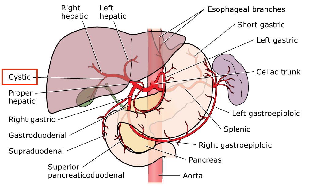
Celiac trunk Splenic left gastric cystic hepatic
95.

During an intense workout, the number of trophic inclusions in the cells of the athlete’s liver and skeletal muscles decreases. What substance belongs to such trophic inclusions?

Explanation

The glycogen granule is essentially an assembly of glucose units (i.e., polymers) that is formed in a branch-like structure via α-glycosidic bonds. As exercise intensity progresses from moderate to high-intensity, muscle glycogenolysis (Glycogenolysis - breakdown of glycogen), liver glycogenolysis and glucose uptake increase such that glucose metabolism predominates. In contrast, there is a reduction in whole body lipid oxidation due to a reduction in both plasma free fatty acids (FFA) and intramuscular triglyceride oxidation.

Starch granules are carbohydrate storage reserves found in plants. 

Melanin found in the skin cortex, protects against UV rays. It has no role in energy production for exercise.

Lipofuscin are yellow-brown pigment granules, considered to be one of the aging or "wear-and-tear" pigments, found in the liver, kidney, heart muscle, retina, adrenals, nerve cells, and ganglion cells.


The waste from broken-down oxalate is called oxalic acid. Oxalic Acid Crystals can be dissolved in water for the effective removal of light stains. It has no role in energy production for exercise.

96. A 49-year-old woman developed a leg edema after a long time spent standing. What is the likely cause of edema development?

Explanation

Edema, (alternate spelling: oedema) is the swelling of the body\'s tissues due to excess interstitial fluid retention. Edema can occur locally, often affecting the extremities (peripheral edema), or generally, affecting the entire body (anasarca).

Fluid moves easily between intravascular and extravascular compartments, and the extent of this movement is determined primarily by the balance between hydrostatic and oncotic pressures.

Hydrostatic pressure refers to the pressure exerted by gravity on a fluid at equilibrium (which is increased when a patient has been standing for a long time), and tends to cause fluid to filter out into the extravascular compartment. Oncotic pressure is a form of osmotic pressure in which proteins in the plasma pull fluid back into the intravascular compartment. The lymphatic system then transports any extra fluid back into the intravascular compartment through the thoracic duct. Therefore, any change in the balance of these pressures that results in a net filtration greater than the lymphatic system can effectively transport can cause edema.

In this case, prolonged standing caused an increased gravitational pull on fluid in the intravascular compartment which increased the hydrostatic pressure resulting in pedal (leg) edema.

97. Autopsy of the body of a 40-year-old patient detected groups of enlarged follicles in the small intestine. Their surface has ridges and fissures arranged in a pattern that resembles gyri and sulci of the brain. The follicles protrude above the surface of the intestinal mucosa. On section they are gray red and juicy. Microscopy shows proliferation of monocytes, histiocytes and reticular cells, there are macrophage clusters that form granulomas, while lymphocytes are depleted. What disease can be characterized by these changes?

Explanation

Enteric/Typhoid fever is a life-threatening illness caused by infection with the bacterium Salmonella enterica serotype Typhi (S. typhi), usually transmitted through food and drinks contaminated with fecal matter. It is associated with symptoms that include high fever, fatigue, headache, abdominal pain, diarrhea or constipation, weight loss, and a rash known as \"rose spots.\" Early diagnosis and treatment are important because serious complications, including severe intestinal bleeding or perforation (due to ulcerations), can develop within a few weeks.

They exhibit ileal mucosal hypertrophy caused by a neutrophil-poor monocyte/macrophage-rich hyperplasia. Though diffuse areas were present, much of the lesional proliferation was nodular, representing macrophage infiltration and colonization by the monocytes and macrophages.

Mucosal hypertrophy was described in this question as -surface has ridges and fissures that resembles gyri and sulci of the brain; follicles protrude above the surface of the intestinal mucosa.

For differential diagnosis on microscopy findings:

S. typhi: primarily monocytes, macrophages

Salmonella - Salmonellosis (except Salmonella typhi): predominantly Neutrophils

Shigella - Dysentery: Primarily Neutrophils

Cholera: B Lymphocytes - antibody secreting cells in Peyer’s patches

Amebiasis: Neutrophils

98. General catabolism pathway of biological macromolecules includes besides tricarbonic acid cycle and mitochondrial respiratory chain, the process of pyruvate oxidative decarboxylation. What is the product of pyruvate oxidative decarboxylation?

Explanation

Pyruvate oxidative decarboxylation produces Acetyl Co-A

Pyruvate non-oxidative decarboxylation produces Lactic acid

In aerobic organisms, glucose and other sugars, fatty acids, and most amino acids are degraded to the Acetyl group of Acetyl-CoA, the form in which the citric acid cycle (Tricyclic acid cycle) accepts most of its fuel input. The oxidative decarboxylation of Pyruvate to form Acetyl-CoA is the link between Glycolysis and the Citric acid cycle. The reaction occurs in the mitochondrial matrix. The pyruvate derived from glucose by glycolysis is dehydrogenated to yield acetyl CoA and CO2 by the enzyme pyruvate dehydrogenase complex.

99. A mountain climber was climbing a mountain for several days. At the altitude of 5000 meters he developed tachypnea, tachycardia and bursting headache. What is the likely cause of these signs?

Explanation

The further you move away from sea level up into higher altitudes, the lower the air pressure is. This can result in hypoxia.

Hypoxia in medicine refers to a condition of the body in which the tissues are starved of oxygen. In its extreme form, where oxygen is entirely absent, the condition is called anoxia.

The percentage of oxygen in the air at two miles (3.2 km/3,200m) is essentially the same as at sea level.

Atmospheric pressure and inspired oxygen pressure fall roughly linearly with altitude to be 50% of the sea level value at 5500 m and only 30% of the sea level value at 8900 m (the height of the summit of Everest) due to the fact that the atmosphere is less dense--that is, the air molecules are farther apart. A fall in inspired oxygen pressure reduces the driving pressure for gas exchange in the lungs and in turn produces a cascade of effects right down to the level of the mitochondria, the final destination of the oxygen.

Hypoxemic type, in which the oxygen pressure in the blood going to the tissues is too low to saturate the hemoglobin. Often encountered in pilots, mountain climbers, and people living at high altitudes—due to reduced partial pressure of oxygen.

100. A patient has heart rhythm disturbances. To restore the rhythm, the doctors prescribed the patient calcium antagonists. What effect do calcium ions have on the myocardium?

Explanation

Now you have to be careful here to know what the question is actually talking about.

The doctor actually prescribed calcium antagonist to the patient but the question is asking you about the effect of Calcium ions on the myocardium.

With this understanding, the question becomes easy. You know calcium helps the muscles contract forcefully and that’s it. Increasing contractility is done primarily through increasing the influx of calcium or maintaining higher calcium levels in the cytosol of cardiac myocytes during an action potential.

If the question asked about the effect of calcium antagonist, then decreased force and frequency of contraction would be the right answer.

101. Regional lymph nodes surrounding an infected wound are enlarged. Histological examination shows increased number of macrophages, lymphocytes and lymphatic follicles in the cortical layer of the lymph nodes, as well as large amount of plasma cells. What process in the lymph nodes is indicated by these histologic changes?

Explanation

Lymph nodes and Peyer's patches play key roles in the development of an appropriate and efficient immune response. Once an Antigen (Ag) is captured by Ag-processing cells, it is rapidly carried to the nearest lymph node, where it is presented to specific lymphocytes to trigger an immune response. During an infection, lymphocyte recruitment from the periphery is enhanced due to a widening of the primary arteriole feeding the lymph node. This leads to the increased number of different types of white blood cells in the lymph node.


In contrast, any form of deficiency, acquired or congenital will be characterized by absent white blood cells in the lymph node. 

Neoplastic changes will be characterized by an increase in only one subtype of white blood cells e.g. a B-cell lymphoma will have an increase in the amount of B cells alone and other types of white blood cells are either normal or decreased.


The type of transplant rejection will determine which cells are increased (e.g. Hyperacute: B Cells, antibodies).

 
102. Autopsy of the body of 6 month old child, who died of sepsis, revealed the absence of thymus and diminished size and weight of the spleen. Microscopy of the spleen detected the absence of periarterial T dependent zones of the follicles and red pulp depletion; In the lymph nodes there is no paracortical zone that is represented mainly by T lymphocytes. B zones in the peripheral immune organs are developed normally. What pathological process is it?

Explanation

DiGeorge syndrome is a congenital immunodeficiency disorder in which the thymus gland is absent or underdeveloped at birth and is most commonly associated with a microdeletion at chromosome 22q11. Children with DiGeorge syndrome are born with several abnormalities, including heart defects, underdeveloped or absent parathyroid glands, an underdeveloped or absent thymus gland, and characteristic facial features.


It is associated with susceptibility to infections (sepsis) due to decreased T cell production and function due to an absent or poorly developed thymus. The thymus is the “school house” where T-cells are educated to fight infection and prevent autoimmunity. T cells are also found in the paracortical zone in the lymph node, so it is expected that the zone will be decreased or absent in the lymphoid tissues.

103. A patient was hospitalized into an infectious diseases department. His skin is dry, with low turgor, stool resembles rice water. The patient was diagnosed with cholera. What water-electrolyte imbalance is likely to occur during this disease?

Explanation

Dehydration (hypohydration, hypohydria, exicosis): in dehydration, the extracellular fluid (ECF) and sodium ions are lost.

*Isoosmolar dehydration: based on proportional volume decrease of fluids and electrolytes. Characterised by isotonic loss of both water and solutes from the extracellular fluid e.g. acute renal failure (stage of polyuria), Diarrhea (especially in cholera - there is equal loss of water and electrolytes), blood loss, burns etc.

*Hypoosmolar dehydration (salt deficit) develops due to diarrhea, vomiting, sweating, adrenal insufficiency etc.


*Hyperosmolar dehydration (water deficit) develops due to the loss of the fluid which lacks electrolytes e.g. in diabetes insipidus, hyperventilation

104. In the enlarged cervical lymph node of a 14 year old girl microscopy detected the following: tissue structure of the lymph node is disturbed, no lymphoid follicles, there are sclerotic areas and necrotic foci, cellular composition of the lymph node is polymorphous, there are lymphocytes, eosinophils, large atypical cells with multilobular nuclei (Berezovsky-Reed_sternberg cells) and large mononuclear cells. Make the diagnosis:

Explanation

Hodgkin lymphoma (also called Hodgkin’s lymphoma, Hodgkin’s disease and lymphogranulomatosis) is a clearly defined malignant disease of the lymphatic system. Hodgkin lymphoma characteristically presents with Hodgkin and Reed-Sternberg cells. When the cells are mononucleated, they are called Hodgkin cells. When they are multinucleated/multilobular, they are called Reed-Sternberg cells.

Classical diagnostic Reed-Sternberg cells are large, have abundant slightly basophilic or amphophilic cytoplasm and have at least two nuclear lobes or nuclei. Diagnostic Reed-Sternberg cells must have at least two nucleoli in two separate nuclear lobes.

Burkitt Lymphoma associated with Ebstein Barr virus (EBV), translocation t(8;14).

Mycosis fungoides/Sezary syndrome: cutaneous T cell lymphoma with skin patches and plaques.

Acute Lymphocytic Leukemia: common in children, associated with Down syndrome

Chronic Lymphocytic Leukemia: associated with Smudge cells (Gumprecht shadows) i.e. crushed or ruptured cells.

105. A 7-year-old girl has signs of anemia. Laboratory testing determined the deficiency of pyruvate kinase in her erythrocytes. In this case the main role in anemia development belongs to the disturbance of a certain process. What process is disturbed in this girl?

Explanation

Pyruvate kinase deficiency is an autosomal recessive condition. Pyruvate Kinase deficiency - decreased ATP production - rigid red blood cells - hemolysis - hemolytic anemia.

Pyruvate kinase is an enzyme involved in glycolysis. The pyruvate kinase enzyme is required for conversion of phosphoenolpyruvate into pyruvate and ATP. This is the final step in Glycolysis which supplies erythrocytes with energy and ATP for cell membrane stability. Lack of this enzyme, therefore, hinders the completion of the reaction with the effect being:

  • Increase in the level of dangerous precursors;

  • Decrease in a number of products such as pyruvate, ATP and lactate.


Pyruvate kinase is not involved in any of the other reactions listed - Peroxide decomposition, oxidative phosphorylation, amino acid deamination, tissue respiration.

106. Biopsy material of a 67-year-old man, who for 17 years has been suffering from chronic bronchitis, shows cylindrical bronchiectasis, the mucous glands have cystic changes, there are patches, where cuboidal epithelium is replaced with stratified squamous epithelium. What pathological process was detected in the bronchial mucosa?

Explanation

Metaplasia is a change of cells to a form that does not normally occur in the tissue in which it is found. In this case, there is a change from a normally occurring cuboidal cells to stratified squamous epithelium which is not normally found in the bronchi.

Metaplasia is the conversion of one adult tissue type into another, related and more durable, tissue type. The most prevalent examples are conversion of fibrous tissue into bone, or columnar mucosal epithelium into stratified squamous epithelium.

Squamous metaplasia is a pre-neoplastic change of the bronchial epithelium observed in the lungs in response to toxic injury induced by cigarette smoke. It is part of a multi-stage process which may eventually lead to full neoplastic transformation, i.e. bronchial carcinoma. Also, Barrett esophagus - esophageal adenocarcinoma is an example.

Hypertrophy is an increase in size of cells e.g. cardiac hypertrophy.

Hyperplasia is an increase in the number of cells e.g. benign prostatic hyperplasia

Heterotopia - ectopic tissue.


Dysplasia: loss of uniformity in cell size and shape (pleomorphism).

107. A 59-year-old man in a severe condition was hospitalized into the cardiology department with the following diagnosis; acute myocardial infarction in the region of the posterior wall of the left ventricle and septum, initial pulmonary edema development is primary in this patient?

Explanation

Pulmonary edema refers to the accumulation of excessive fluid in the alveolar walls and alveolar spaces of the lungs. Pulmonary edema is often caused by congestive heart failure. When the heart is not able to pump efficiently, blood can back up into the veins that take blood through the lungs.

As the pressure in these blood vessels increases, fluid is pushed into the air spaces (alveoli) in the lungs. This fluid reduces normal oxygen movement through the lungs. These two factors combine to cause shortness of breath.

All the factors which contribute to increased pressure in the left side and pooling of blood on the left side of the heart can cause cardiogenic pulmonary edema. The result of all these conditions will be increased pressure on the left side of the heart: increased pulmonary venous pressure--> increased capillary pressure in lungs--> pulmonary edema.

  • Coronary artery disease with left ventricular failure (myocardial infarction)

  • Congestive heart failure

  • Cardiomyopathy

  • Valvular heart diseases on the left side of the heart (stenosis and regurgitation)

  • Cardiac arrhythmias

108. A patient developed a purulent inflammatory process in the periodontal tissues. The process was caused by activation of the microorganisms inherent in the body, which are a part of oral mucosal microflora. What type of infection is it?

Explanation

Autoinfection is an infection by a pathogenic agent already within the body or infection transferred from one part of the body to another. With this definition, we can break down the question and arrive at an answer.

The purulent inflammatory process in the periodontal tissues was caused by a microorganism which is a part of the normal oral mucosa. Periodontal disease, also known as gum disease, is a set of inflammatory conditions affecting the tissues surrounding the teeth. So, the pathogenic agent is already within the body, in the oral cavity. 

In short, the microorganism didn’t come from outside the body, its part of the normal oral microflora. So, it is a case of self-infection.

Superinfection is infection occurring after or on top of an earlier infection e.g. Hepatitis B patient superinfected by Hepatitis D.

Reinfection: infection by the same organism following an earlier infection.

Relapse is a case of deterioration in someone's state of health after a temporary improvement.


Exogenous (outside): Exogenous infections involve a pathogen entering a patient's body from their environment. These pathogens can be introduced through a contaminated device, healthcare worker, surface, or other vector.

109. A patient suffers from disturbed renal function. To check the filtration ability of the kidneys, he was referred for clearance measurement of the following substance:

Explanation

Creatinine is a waste product formed by the normal breakdown of muscle cells. Healthy kidneys take creatinine out of the blood and put it into the urine to leave the body. When the kidneys are not working well, creatinine builds up in the blood. 

The biochemical marker creatinine found in serum and urine is commonly used in the estimation of Glomerular Filtration Rate (GFR). Creatinine clearance is the volume of blood plasma cleared of creatinine per unit time. It is a rapid and cost-effective method for the measurement of renal function. Both Creatinine clearance and Glomerular Filtration Rate can be measured using the comparative values of creatinine in blood and urine.


Creatinine is commonly used as a measure of kidney function. The creatinine clearance test is used to monitor the progression of renal disease. The diagnosis of renal failure is usually suspected when serum creatinine is greater than the upper limit of the “normal” interval.

110. During emotional excitation the heart rate of a 30-year-old person reached 112/min. The increased heart rate was caused by a change that occurred in a certain structure of the cardiac conduction system. Name this structure:

Explanation

The sinoatrial (SA) node is a small strip of modified cardiac muscle, situated in the superior part of lateral wall of right atrium, just below the opening of superior vena cava. The fibers of this node do not have contractile elements. SA node is called the pacemaker because the rate of production of impulse (rhythmicity) is higher in SA node than in other parts. The rate is 60-100/min and higher during tachycardia when stimulated by sympathetic effects. In this case, emotional excitation led to the tachycardia (112/min) observed. Atrioventricular (AV) node is 40-60/min. Purkinje fibers: 20 - 40/min His bundle: 20 or less per min.
111.

A 40-year-old man with pulmonary tuberculosis was prescribed isoniazid. Prolonged taking of this drug can result in the development of the following vitamin deficiency:

Explanation

Isoniazid (INH) is a potent bactericidal antibiotic used in the treatment of tuberculosis. Treatment with Isoniazid therapy has a risk of toxicity, which can be acute or chronic. Acute toxicity manifests as neurological symptoms; while chronic toxicity usually presents as hepatotoxicity and peripheral neuropathy. 

Isoniazid causes peripheral neuropathy by 2 different mechanisms. First, Isoniazid metabolites directly inactivate pyridoxine species. It also acts by inhibiting the enzyme pyridoxine phosphokinase which is a necessary enzyme to convert pyridoxine to its active form of pyridoxal 5' phosphate which is a very important cofactor in many reactions. 

The pyridoxine deficiency induced by the use of Isoniazid leads to reduced production of GABA, as it is usually a product of pyridoxine-dependent decarboxylation reaction. Thus GABA deficiency can manifest as seizures especially in the acute setting of toxicity.


Vitamin B6 (pyridoxine) supplementation during isoniazid (INH) therapy is necessary in some patients to prevent the development of peripheral neuropathy.

112. A patient with suppurative bronchitis was hospitalized into the pulmonology department. As a part of complex therapy, he was prescribed a medicine that liquefies sputum and facilitates expectoration. Name this medicine:

Explanation

Acetylcysteine inhalation is used along with other treatments to relieve chest congestion due to thick or abnormal mucous secretions in people with lung conditions. It is mainly used in thinning mucus and to treat Paracetamol toxicity.

When inhaled by mouth, acetylcysteine is used to help thin and loosen mucus in the airways due to certain lung diseases (such as emphysema, bronchitis, cystic fibrosis, pneumonia). This effect helps you to clear the mucus from your lungs so that you can breathe easier. 


When taken by mouth, acetylcysteine is used to prevent liver damage from acetaminophen (paracetamol) overdose.

113. In an experiment on an isolated squid giant axon submerged in a salt solution, the extracellular potassium ions concentration was increased to the level of the intracellular potassium ions concentration. What changes in the membrane potential will occur in this case?

Explanation

Membrane potential is what we use to describe the difference in voltage (or electrical potential) between the inside and outside of a cell.

Membrane potential refers to the difference in charge between the inside and outside of a neuron, which is created due to the unequal distribution of ions on both sides of the cell.

For a resting membrane potential, there is more potassium ions within the cell (intracellular) than outside the cell (extracellular). This unequal distribution of ions is what drives the movement of ions and this movement of ions gives rise to what is called an action potential. However, in a situation when we have equal charges on both sides; therefore the membrane has no potential. (extracellular Potassium equals Intracellular Potassium). 


Ions can’t move in or out of the cell because they have equal charges (equal number of ions) - the membrane potential is non-existent in this case. SO increasing the extracellular potassium ion to equal the intracellular ion will make the membrane potential disappear.

114. A patient with essential hypertension was prescribed hydrochlorothiazide as a part of complex therapy. What mechanism of drug action facilitates a decrease in blood pressure in this case?

Explanation


Hydrochlorothiazide (HCTZ) is a thiazide-type diuretic which has been widely used to treat hypertension globally. Hydrochlorothiazide inhibits sodium chloride transport in the distal convoluted tubule. More sodium is then excreted in the kidney with accompanying fluid. Pharmacological effects begin in about 2 hours after an oral dose, peaks in 4 hours, and lasts for about 6 to 12 hours. When administered acutely the drug does lower blood pressure by promoting diuresis and decreasing plasma volume.

115. A patient is diagnosed with glucocerebroside lipidosis (Gaucher’s disease) that manifests as splenomegaly, liver enlargement, affected bone tissue and neuropathies. What enzyme of complex lipid catabolism is deficient, causing this disease?

Explanation

Gaucher disease is a rare genetic disorder characterized by the deposition of glucocerebroside in cells of the macrophage-monocyte system. The disorder results from the deficiency of the enzyme glucocerebrosidase. It is an autosomal recessive disorder. Characterized by hepatosplenomegaly, pancytopenia, osteoporosis, avascular necrosis of femur, bone crises.

Niemann-Pick disease: deficiency of Sphingomyelinase resulting in accumulation of Sphingomyelin.

Tay-Sachs disease: deficiency of Hexosaminidase A resulting in accumulation of GM2 ganglioside.

Deficiency of beta-galactosidase leads to the accumulation of sphingolipid intermediates in lysosomes of neuronal tissue, resulting in the CNS deterioration typical of GM1. Deficiency of this enzyme also leads to accumulation of the glycosaminoglycan (GAG) keratan sulfate in cartilage which is suspected to cause the skeletal findings associated with mucopolysaccharidosis type IVB (MPS IVB).


Hyaluronidase deficiency is a condition caused by mutations in HYAL1, and characterized by multiple soft-tissue masses. A rare form of mucopolysaccharidosis characterized by abnormal storage of hyaluronan in lysosomes due to deficiency of hyaluronidase 1.

116. A patient with Parkinson’s disease made an appointment with the doctor. The doctor prescribed him a medicine that is a dopamine precursor in the central nervous system. What medicine of those listed below has such mechanism of action?

Explanation

Levodopa is the precursor to dopamine. Most commonly, clinicians use levodopa as a dopamine replacement agent for the treatment of Parkinson's disease. It is most effectively used to control bradykinetic symptoms that are apparent in Parkinson's disease (PD) and it is the most effective medication to improve the quality of life in patients with idiopathic PD.

Degeneration of the substantia nigra occurs in patients with Parkinson's disease decreasing the striatal dopamine levels. Levodopa converts to dopamine in both the CNS and periphery. In order to increase the bioavailability of levodopa and decrease its side effects, it is often administered in combination with peripheral decarboxylase inhibitors (such as carbidopa and benserazide).

Platyphyllin hydrotartrate used to treat intestinal colic.

Methacin is an analgesic (pain killer).

Lorazepam is an anticonvulsant and anxiolytic from the class of drugs called benzodiazepines.


Ipratopium bromide is used to control and prevent symptoms (wheezing and shortness of breath) caused by ongoing lung disease (chronic obstructive pulmonary disease-COPD which includes bronchitis and emphysema). It works by relaxing the muscles around the airways so that they open up and you can breathe more easily.

117. During gastric resection the patient received mixed anesthesia with tubocurarine chloride muscle relaxant. To restore unassisted respiration in the patient, the patient was given proserin. What pharmacological group does this drug belong to?

Explanation

Most uses of cholinesterase inhibitors are based on a common mechanism of action initiated by inhibition of acetylcholinesterase (AChE). Extensive inhibition of this enzyme leads to accumulation of the neurotransmitter acetylcholine and enhanced stimulation of postsynaptic cholinergic receptors. This action is beneficial in cases where a reduction in cholinergic transmission contributes to clinical symptoms, e.g., low muscle tone - to reverse the effects of muscle relaxants such as gallamine and tubocurarine.

Proserin or Neostigmine is a parasympathomimetic, specifically, a reversible cholinesterase inhibitor. The drug inhibits acetylcholinesterase which is responsible for the degradation of acetylcholine. So, with acetylcholinesterase inhibited, more acetylcholine is present By interfering with the breakdown of acetylcholine, neostigmine indirectly stimulates both nicotinic and muscarinic receptors which are involved in muscle contraction.. It does not cross the blood-brain barrier.

Neuromuscular junctions use Nicotinic receptors. Muscarinic receptors are not present in Neuromuscular junctions. So any class of drugs affecting only Muscarinic receptors will not be effective in this case.

Calcium channel blockers will further worsen the condition as muscles require Calcium for adequate contraction.


Angiotensin converting Enzyme Inhibitors are used in hypertensive patients.

118. A patient has a trauma of the knee joint with crushed patella. With such injury, it is likely that the tendon of a certain thigh muscle is damaged. Name this muscle:

Explanation

The quadriceps femoris is a hip flexor and a knee extensor. It consists of four individual muscles; three vastus muscles (vastus medialis, lateralis and Intermedius) and the rectus femoris. They form the main bulk of the thigh, and collectively are one of the most powerful muscles in the body. It is located in the anterior compartment of the thigh.


The quadriceps tendon is located just above the kneecap (patella) and connects the quadriceps muscles in the front of the thigh to the top of the kneecap. The patellar tendon is located just below the kneecap. It connects the kneecap to the shinbone (tibia). The function of the quadriceps tendon and patellar tendon is to work with the muscles in the front of the thigh to straighten the knee.

Quadriceps Muscle Tendon Patella

119. Mitochondrial destruction is observed in some hereditary diseases (for example, Kearns-Sayre syndrome). What processes can be disturbed, as a result, in the cell?

Explanation

Most of the adenosine triphosphate (ATP) synthesized during glucose metabolism is produced in the mitochondria through oxidative phosphorylation. This is a complex reaction powered by the proton gradient across the mitochondrial inner membrane, which is generated by mitochondrial respiration.


Kearns-Sayre syndrome (KSS) is caused by deletions of large portions of mitochondrial DNA (mtDNA), resulting in the loss of genes involved in the oxidative phosphorylation pathway. With this mutation that affects oxidative phosphorylation, it is obvious that this will lead to less ATP synthesis in the mitochondria.

120. A 36-year-old man developed angina pectoris attacks immediately after a past case of staphylococcal sepsis. Coronarography detected mural thrombosis without signs of atherosclerosis in the left coronary artery. Thrombus formation occurred in the result of damage to the vascular endothelium and release of:

Explanation

Injury to endothelium is accompanied by loss of protective molecules and expression of adhesive molecules, procoagulant activities, and mitogenic factors, leading to development of thrombosis. Platelet activating factor is one of the procoagulant molecules released when endothelial cells are damaged.


Platelet activating factor (PAF) is an endogenous phospholipid mediator of inflammation that is produced by inflammatory, endothelial, and lamina propria cells. It is involved in platelet activation and aggregation after antigen challenge (Staphylococcal infection) or endothelial injury.

121. T lymphocytes were affected by HIV. In the process, viral enzyme reverse transcriptase (RNA-dependent DNA-polymerase) catalyzed the synthesis of:

Explanation

HIV is a RNA virus from the family Retroviridae. One of its unique properties is the presence of an enzyme known as reverse transcriptase.

I’ll explain this in very simple terms instead of using biochemical terms that might be confusing.

HIV is a RNA virus and the human genome has DNA. When HIV infects human cells, its RNA virus is usually converted into DNA so that the DNA can be incorporated into the human DNA. Once this is done, then the human cells help the HIV to replicate.

HIV enters the cell as RNA virus.

The RNA is Converted by Reverse transcriptase to DNA (RNA dependent-DNA polymerase)

This DNA is then incorporated into the Human DNA using the enzyme Integrase

Once the HIV DNA is integrated, when the human cells want to replicate they also replicate the HIV DNA without knowing.

This is how the HIV replicates using the human cells.

In conclusion, Reverse Transcriptase synthesizes DNA using the viral (HIV) RNA.

HIV replication Reverse transcription

122. There is a large amount of glucose oxidation metabolites dissolved in the cytoplasm of myocytes. Name one such metabolite that converts directly into lactate:

Explanation

When tissue can’t be supplied by oxygen, or during exercise when less oxygen reaches muscle than their need, then pyruvate acts as a terminal electron acceptor from NADH (formed during glycolysis) and converted into lactate, a process called lactic acid fermentation. RBC, retina cells, and muscles during exercise and during hypoxic condition respire by lactic acid fermentation.

Lactate formed in the active muscles is transported to the liver where it can be broken down or restored into glucose, the restored glucose from lactate is transported to muscles - this cycle is called a Cori cycle.


Of all the options listed, only Pyruvate converts directly into lactate under anaerobic conditions. Pyruvate can be converted directly to Acetyl-CoA under aerobic conditions.

123. From the feces of a patient with acute gastroenteritis a pure culture of microorganisms was obtained. The microorganisms are small mobile slightly curved gram-negative bacilli that within 6 hours grow into a light blue film on the 1% alkaline peptone water. Such properties are characteristic of the following microorganisms:

Explanation

Vibrio cholerae is a Gram-negative enteric pathogen. It is flagellated, comma shaped, oxidase +, grows in alkaline media and produces profuse rice-water diarrhea.


Although V. cholerae will grow on a variety of commonly used agar media, isolation from fecal specimens is more easily accomplished with specialized media. Alkaline peptone water (APW) is recommended as an enrichment broth, and thiosulfate citrate bile salts sucrose (TCBS) agar is the selective agar medium of choice for isolating V. cholerae.

124. A bioterrorist mailed an envelope with a powder that is suspected to contain anthrax causative agent. This envelope can remain dangerous for a long time, because anthrax causative agent:

Explanation

Bacillus anthracis (B. anthracis), the etiologic agent of anthrax is a Gram positive, spore forming, non-motile bacterium. This is supposed to be one of the most potent Biological Weapon agents because its spores are extremely resistant to natural conditions and can survive for several decades in the environment. B. anthracis spores enter the body through skin lesion (cutaneous anthrax), lungs (pulmonary anthrax), or gastrointestinal route (gastrointestinal anthrax) and germinate, giving rise to the vegetative form.

Due to the fact that the spores can survive for a long time, this property makes it a dangerous bioterrorism weapon.

Spores of Bacillus genus are known to have a half life of about 100 years. Spores are dormant form of the bacterium which returns into vegetative form on receiving the signals for germination. The surprisingly resistant spores have earned the status of potential bioterror weapon for anthrax.

125.

A 65-year-old woman against the background of chronic heart failure developed secondary hyperaldosteronism. What medicine should be prescribed to increase the patient’s diuresis?

Explanation

What this question wants from you is to know the diuretic that acts as an antagonist to Aldosterone because this patient has developed secondary hyperaldosteronism.

In untreated congestive heart failure, aldosterone plasma concentrations are elevated in proportion to the severity of the disease and are further increased by the use of diuretic treatment. 

The Renin-Angiotensin-Aldosterone system (RAAS) is the complex mechanism by which the kidneys control the cardiovascular system. The system goes awry in patients with heart failure; in these persons, the decreased renal blood flow is not due to low blood volume but to a heart pump that is not working efficiently. The moment there is decreased blood flow to the kidney, RAAS is activated resulting in an increased Aldosterone level - (secondary hyperaldosteronism).


Spironolactone is a specialized antagonist of aldosterone. It acts as a competitive binding agent at the aldosterone receptor site in the distal convoluted renal tubules, preventing the formation of a protein important in the sodium-potassium exchange in the kidneys. This action causes increased amounts of water and sodium to be excreted while potassium is conserved.

126. When examining a biopsy material obtained from the thyroid gland, the pathologist discovered lymphocyte infiltration of the thyroid tissues and destruction of the parenchymal elements. Diffuse lymphocyte infiltration with lymphoid follicles was detected in the stroma. What is the most likely diagnosis?

Explanation

Hashimoto’s thyroiditis is an autoimmune inflammatory disorder of the thyroid gland. It is also referred to as chronic lymphocytic thyroiditis or chronic autoimmune thyroiditis. The synonyms of this disease are derived from its pathological features which include diffuse lymphocytic infiltration of the thyroid gland, first described by Hashimoto in 1912, and the presence of autoantibodies against thyroid antigens. It is the most common cause of hypothyroidism in the developed world and is more prevalent among women.

Hashimoto’s thyroiditis is defined with the histologic features of diffuse lymphocytic thyroiditis with follicular atrophy, diffuse destruction of thyroid follicles, fibrosis, and follicular cell regeneration.


Graves' Disease is a type of hyperthyroidism that is due to the production of autoantibodies against the TSH receptor on the follicular epithelial cells. These antibodies mimic the effects of TSH and cause overproduction and release of thyroid hormone.

127. A patient used an indirect-acting adrenergic agonist to treat rhinitis. After the patient has been putting in the nose drops for several days, the casoconstrictive effect of the drug gradually diminished. Name this phenomenon:

Explanation

Tachyphylaxis: rapidly diminishing response to successive doses of a drug, rendering it less effective. It is the continued or repeated exposure to a drug that may lead to a weakened pharmacological response. This is presumed to be a consequence of diminished receptor sensitivity in response to consistent stimulation by a drug agonist, which produces a diminished pharmacological response in consequence.

In this case, the vasoconstrictive effect of the indirect-acting adrenergic agonist gradually diminished after several days.

Teratogens are substances that may cause birth defects via a toxic effect on an embryo or fetus. Known teratogens include: thalidomide, mercury, alcohol, lead etc.

An allergy is an immune system response to a foreign substance that's not typically harmful to your body. These foreign substances are called allergens. They can include certain foods, pollen, or pet dander.

Idiosyncrasy is an abnormal physical reaction by an individual to a food or drug. This is unique to the patient.


Drug cumulation is the process by which blood levels of a drug build-up, thereby increasing its therapeutic and toxic effects.

128. A 40-year-old woman on examination presents with intensified basal metabolic rate. What hormone present in excess leads to such condition?

Explanation

Thyroid hormone controls the basal metabolic rate. A decreased basal metabolic rate indicates hypothyroidism and an increased (intensified) basal metabolic rate indicates Hyperthyroidism.

The major thyroid hormone secreted by the thyroid gland is thyroxine, also called T4 because it contains four iodine atoms. To exert its effects, T4 is converted to triiodothyronine (T3) by the removal of an iodine atom. This occurs mainly in the liver and in certain tissues where T3 acts, such as in the brain. 

In Hyperthyroidism, it is expected that Thyroxine (T4) and Triiodothyronine (T3) levels will be present in excess.


The other hormones listed don't control the basal metabolic rate.

129. Among lymphocytes there is a population of cells that have membrane receptors to IgM, activate in response to certain antigens, reproduce mitotically, differentiate into plasma cells that product antibodies (immunoglobulins). Name these cells:

Explanation

Immune responses have been classified as cellular or humoral. Cellular responses are mediated by T lymphocytes, which recognize and attack their targets directly or indirectly by enlisting the help of other immune cells, while humoral responses are characterized by the production of antibodies by B lymphocytes and their progeny, plasma cells. 

When B cells are stimulated, B cells differentiate into plasma cells that produce antibody molecules closely modeled after the receptors of the precursor B cell. Once released into the blood and lymph, these antibody molecules bind to the target antigen (foreign substance) and initiate its neutralization or destruction.


Antibodies, also known as immunoglobulins, are glycosylated protein molecules present on the surface of B cells (surface immunoglobulins) serving as antigen receptors (BCR), or are secreted into the extracellular space where they can bind and neutralize their target antigens. Five isotypes, or classes, of antibodies (IgM, IgD, IgG, IgA, and IgE) exist.

130. The ophthalmologist noticed a purulent discharge from the conjunctiva of a newborn. Microscopy of the smear obtained from the conjunctiva found three a large number of leukocytes, as well as gram-negative bean-shaped diplococci located inside the leukocytes. What is the causative agent of this disease?

Explanation

Neisseria gonorrhoeae is a gram negative (Gram -) diplococci. It is often intracellular within Neutrophils. Often transmitted sexually or perinatally. 

This question is a classic vignette of neonatal conjunctivitis which usually presents 2 - 5 days after birth.


Women with untreated gonorrhea can pass the bacteria to her baby during childbirth. Neonatal conjunctivitis, also called ophthalmia neonatorum, typically presents during the first four weeks of life. The infection is usually acquired during delivery and is the most common ocular disease in neonates. Typical symptoms are persistent tearing and a mucoid discharge in the inner corner of the eye, red eyes, and swelling of the eyelids.

131.

A biopsy material was obtained from the arches of the patient’s soft palate due to a suspected tumor (Macroscopy detected an ulcer with the dense floor). In the biopsy material the following was detected: necrosis of the mucosa with infiltration of the submucosal layer by lymphocytes, epithelioid cells, plasma cells and single neutrophils. Notable is the presence of marked endovasculitis and perivasculitis. The described changes are characteristic of:

Explanation

Syphilis is caused by Treponema pallidum. The microscopic findings described in this question is consistent with findings associated with Syphilis. 

The syphilis granuloma consists of a central area of fibrinoid or caseous necrosis surrounded by mononuclear inflammatory cells, mostly plasma cells, lymphocytes, epithelioid cells, and seldom giant Langhans cells. Surrounding it is the granulations tissues, endovasculitis and perivasculitis.

 
132. When examining a child, the pediatrician noted that the child presents with delayed physical and mental development. Urinalysis showed an acute increase in the levels of a keto acid that produce qualitative color reaction with ferric chloride. What metabolic disturbance was detected in this case?

Explanation

Phenylketonuria (PKU) is an autosomal recessive inborn error of phenylalanine metabolism resulting from deficiency of phenylalanine hydroxylase. Untreated PKU is associated with an abnormal phenotype which includes growth failure, poor skin pigmentation, microcephaly, seizures, global developmental delay and severe intellectual impairment. 

When this enzyme is deficient, abnormally high levels of phenylalanine accumulate in the blood and other tissue fluids, and it is the excess of phenylalanine which is held to be responsible, directly or indirectly, for the progressive cerebral damage in the untreated patient. Some of the phenylalanine is excreted unchanged in the urine and the remainder as a variety of breakdown products. Among them is phenylpyruvic acid, and this causes the characteristic greenish-blue colour reaction when a few drops of a solution of ferric chloride are added to the urine. 

Alkaptonuria: enzyme deficiency is homogentisic dioxygenase (HGD), which is used to break down a toxic substance called homogentisic acid. When there is a deficiency of this enzyme, homogentisic acid builds up in your body. Affected individuals may have dark urine or urine that turns black when exposed to air - this is not a ferric chloride reaction. (Phenylketonuria is involved in colour change on exposure to ferric chloride, not Alkaptonuria).

Albinism: Enzyme deficiency is Tyrosinase, which synthesizes melanin from the amino acid tyrosine.

Tyrosinemia: enzyme deficiency is fumarylacetoacetate hydrolase (FAH), which is needed for the final breakdown of the amino acid tyrosine.


Cystinuria is an inherited disease that causes stones made of the amino acid cystine to form in the kidneys, bladder, and ureters. The mutations result in the abnormal transport of cystine in the kidney and this leads to the symptoms of cystinuria.
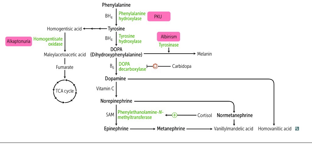
Phenylalanine Phenylketonuria Alkaptonuria Vanillylmandelic acid Homovanillic acid

133. The patient’s ECG shows that in the second standard lead from the extremities the P waves are positive, their amplitude is 0.1 mV (norm is 0.05-0.25 mV), duration – 0.1 seconds (norm is 0.07-0.10 seconds). It can be concluded that the following process occurs normally in the cardiac atria:

Explanation

From the values given in the question:

P wave amplitude 0.1mV (norm: 0.05 - 0.25mV) - this value falls within normal range.

P wave duration 0.1secs (norm: 0.07 - 0.10secs) - this value falls within normal range.

Also, we know that the P wave represents Atrial depolarization.

QRS complex represents Ventricular depolarization.


T wave represents Ventricular repolarization.

[caption id="attachment_1118" align="aligncenter" width="728"] P-wave indicates Atrial depolarization, QRS complex- ventricular depolarization, T-wave- ventricular repolarization.[/caption]

134.

Some adults develop signs of dyspepsia after drinking milk. What enzyme deficiency is associated with milk intolerance?

Explanation

People with lactose intolerance are unable to fully digest the sugar (lactose) in milk. As a result, they have diarrhea, gas and bloating after eating or drinking dairy products. The condition, which is also called lactose malabsorption, is usually harmless, but its symptoms can be uncomfortable.

Lactose intolerance is usually the result of your body not producing enough lactase. Lactase is an enzyme (a protein that causes a chemical reaction to occur) normally produced in your small intestine that's used to digest lactose. If you have a lactase deficiency, it means your body does not produce enough lactase.

Maltase, enzyme that catalyzes the hydrolysis of the disaccharide maltose to the simple sugar glucose. 

Lipase breaks down fats (lipid).

Peptidase breaks down peptide bonds between amino acids in protein.


Amylase breaks down starch into simple sugars.

135. A young woman, a foreign student from Tehran, has made an appointment with the urologist. She complains of the sensation of heaviness in her lower abdomen and a small amount of blood being excreted with urine at the end of each urination. Microscopy of urine detects the presence of parasite eggs, approximately 140×70 micron in size, with a terminal spike. What diagnosis can be made by the infectious disease specialist?

Explanation

Schistosomiasis or snail fever is an endemic parasitic infection caused by various trematodes (flatworm) of the genus Schistosoma. People acquire the disease through contact with water containing infected snails. It is one of the most widespread human parasitic infections in tropical and subtropical regions of the world such as Africa, South America, the Middle East, Asia and the Caribbean.

Bladder schistosomiasis is caused by the Schistosoma haematobium species. The larvae (cercariae), which are released from freshwater snails (intermediate host) into water, penetrate through the human skin. Once the larvae mature into adult worms, they travel into pelvic veins, and eggs are deposited along the bladder wall vessels and are excreted in urine. Symptoms are nonspecific and include suprapubic pain, terminal haematuria and dysuria. Urine cytology reveals microscopic or macroscopic haematuria. A definitive diagnosis is made by urine microscopy of a midday urine sample by demonstrating the eggs, which is oval in shape and has a spiked end.
136. Examination of a patient, who for a long time was taking glucocorticoids, detected lymphopenia. How can the functional state of the patient’s immune system be characterized?

Explanation

Secondary immunodeficiencies are far more common than primary immunodeficiencies. Primary immunodeficiencies are caused by genetic defects affecting cells of the immune system. Secondary or acquired immunodeficiencies arise from a number of conditions, such as treatment with glucocorticoids and immunomodulatory drugs, surgery and trauma, extreme environmental conditions, and chronic infections, such as those caused by HIV.

Glucocorticoids have a strong immunosuppressive, anti-inflammatory and anti-allergic effects on immune cells, tissues and organs. In general, glucocorticoids inhibit leukocyte traffic and thereby the access of leukocytes to the site of inflammation. Furthermore, glucocorticoids interfere with immune cell function and suppress the production and actions of humoral factors involved in the inflammatory process. 


Lymphopenia occurs in response to endogenous or exogenous glucocorticoids. This appears to result in part from the sequestration of lymphocytes in lymphoid organs, including bone marrow. Glucocorticoids also potentiate apoptosis of sensitive lymphocytes.

137.

Autopsy of the body of a 1.5 year old child revealed hemorrhagic skin rashes, moderate hyperemia and edema of the nasopharyngeal mucosa, small hemorrhages in the mucosa and internal organs, markedly dystrophic changes in the liver and myocardium, acute necrotic nephrosis and massive hemorrhages in the adrenal glands. These changes are characteristic of the following disease:

Explanation

Meningococcal infection is an acute infectious process which has three main forms: nasopharyngitis, purulent meningitis and meningococcemia.  This is characterized by periodic epidemics, the disease is more common in children under 5 years, but the disease may occur in persons of any age.

Meningococcal nasopharyngitis is characterized by catarrhal inflammation of the mucosa with marked hyperemia, edema of the posterior wall of the pharynx and hyperplasia of lymphatic follicles. 


Meningococcemia: Changes on the organs are characterized by generalized damage of microcirculation, skin rash, changes in the joints, vascular membrane of the eyes, pericardium, adrenal glands and kidneys. The rash is hemorrhagic, star-like, located mainly on the buttocks and lower extremities. Focal necrosis and hemorrhages or bilateral massive hemorrhages with the development of acute adrenal insufficiency (Waterhouse Friderichsen syndrome) are noted in the adrenals.  Necrosis of nephrothelium of the tubules (necrotic nephrosis) is observed in the kidneys. The death of the patients is caused by bacterial shock, its severity is aggravated by hemorrhages to the adrenals; acute renal insufficiency is not so common (in the adults).

138.

Immediately after moving from horizontal to vertical position, the heart rate of a 23-year-old man increased by 15 beats per minute, his systolic pressure remained unchanged, while his diastolic pressure increased by 10 mmHg. What reflex response of the executive structures caused this increase in the diastolic pressure?

Explanation

The vignette described is talking about changes in venous blood return to the heart.

When a person changes position from a horizontal (lying flat) to vertical (standing) position, there is a transient decrease in blood going to the heart. Most of the blood will pull to the capacitance vessels (veins) in the lower extremities.

This decrease in venous blood return to the heart activates the sympathetic nervous system to constrict the blood vessels, increase heart rate and heart contractility in a quest to increase blood pressure.

Resistance vessels are majorly the arterioles where the alpha 1 receptors are located. Constriction of these vessels tends to increase the blood pressure. Diastolic blood pressure depends on the speed of blood flow and therefore on the total peripheral resistance. This attests to the resistance provided by vessels to blood flow. It is a good indicator of arterial wall elasticity. Constriction of the resistance vessels (arterioles) is the major determinant of diastolic blood pressure.

Capacitance vessels are majorly the veins and constriction of these vessels increase venous blood return to the heart.

139. The patient’s ECG was obtained. What element of ECG allows the doctor to assess the spread of atrial depolarization processes?

Explanation

An electrocardiogram, or ECG, is a recording of the heart’s electrical activity as a graph over a period of time. A typical ECG tracing of the cardiac cycle (heartbeat) consists of a P wave (atrial depolarization), a QRS complex (ventricular depolarization), and a T wave (ventricular repolarization). 


The first wave on an ECG is the P wave, indicating atrial depolarization in which the atria contract (atrial systole). The QRS complex refers to the combination of the Q, R, and S waves, and indicates ventricular depolarization and contraction (ventricular systole).The T Wave indicates ventricular repolarization, in which the ventricles relax following depolarization and contraction.

[caption id="attachment_1118" align="aligncenter" width="728"] P-wave indicates Atrial depolarization, QRS complex- ventricular depolarization, T-wave- ventricular repolarization.[/caption]
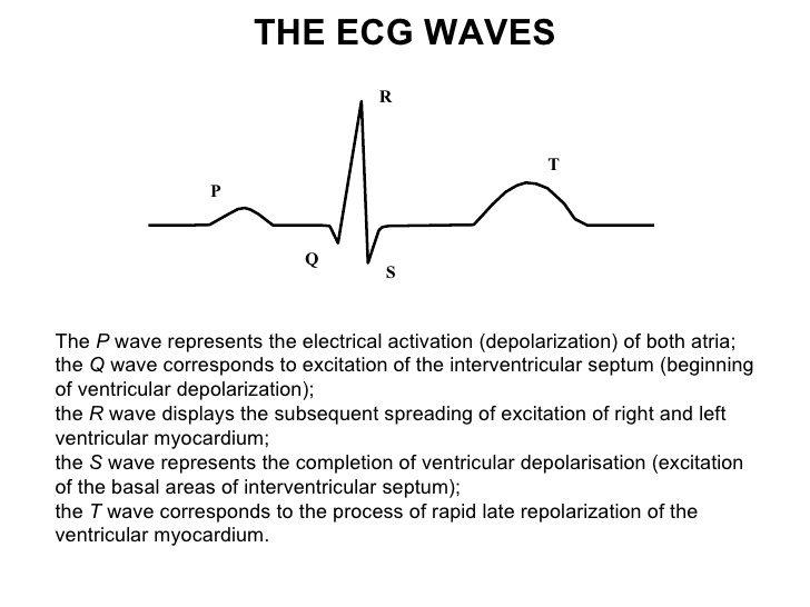
140. A 32-year-old man has tall stature, gynecomastia, female pattern of hair distribution, high pitched voice, delayed mental development and sterility. He was provisionally diagnosed with Klinefelter syndrome. To clarify this diagnosis, it is necessary to analyze the patient’s:

Explanation

Klinefelter syndrome is a genetic condition that results when a boy is born with an extra copy of the X chromosome (XXY). Klinefelter syndrome is a genetic condition affecting males, and it often isn't diagnosed until adulthood.

The main tests used to diagnose Klinefelter syndrome are:

  • Hormone testing. Blood or urine samples can reveal abnormal hormone levels that are a sign of Klinefelter syndrome.

  • Chromosome analysis. Also called karyotype analysis, this test is used to confirm a diagnosis of Klinefelter syndrome. A blood sample is sent to the lab to check the shape and number of chromosomes.


Karyotype analysis or chromosome analysis is used to confirm the diagnosis.

141. A patient with myocardial infarction has acute heart failure. Among the drugs that increases the force of heart contractions the least dangerous in this case will be:

Explanation

Dobutamine is a catecholamine with β-1 and β-2 adrenergic agonist properties which help improve myocardial contractility. In patients with cardiogenic shock due to decompensated heart failure, dobutamine decreases left ventricular end-diastolic pressure and raises blood pressure by increasing cardiac output. It does all these with little effect on heart rate or systemic arterial pressure.

Adrenaline (Epinephrine) can increase the heart rate, contractility and increase blood flow to the heart. However, it also causes constriction of small blood vessels which can reduce blood flow to other organs, including the brain, and may lead to neurological damage.

Caffeine can act on enzymes in the heart that stimulate the intensity of the heart's contractions. Caffeine can facilitate the release of natural hormones that act on the heart to release norepinephrine, which can produce a stimulated effect similar to that of adrenaline.

Aminophylline can cause increased or rapid heart rate, irregular heartbeat, seizures, and skin rash.

Isoprenaline has been shown to cause impairment in perfusion of the subendocardial myocardium resulting in isoprenaline-induced myocardial necrosis.


Of all the answer choices given, Dobutamine is the best and the drug with the least adverse effect profile.  Epinephrine and other vasoconstrictors (caffeine, Isoprenaline) are strictly contraindicated for patients recovering from myocardial infarction supported by the fact that after a myocardial infarction, higher risk of reinfarction is reported.

142. To treat the burns, a patient was prescribed a drug with antiseptic properties that are based on formation of atomic oxygen in the presence of organic substances. This drug has also an astringent(anti-inflammatory) effect due to formation of albuminates. Name this drug:

Explanation

Potassium permanganate (KMnO4) is commercially prepared by mixing solution of potassium hydroxide (KOH) and powdered manganese oxide, with oxidizing agents like potassium chlorate. The mixture is boiled and evaporated and the residue is heated in iron pans until it has acquired a pasty consistency. It is odourless. An aqueous solution with a sweetish astringent taste. It is water-soluble and more soluble in boiling water.

This compound is used as a disinfectant to cure certain skin conditions like foot fungal infections, dermatitis. Another important application of potassium permanganate is in the treatment of bacterial infections. It is also used as a bleaching agent, as a pesticide, and as an antiseptic.

2KMnO4 + H2O = 2KOH + 2MnO2 + 3O2

Potassium permanganate liberates oxygen in atomic form.

143. A man with caisson disease died with the signs of acute disturbances of cerebral circulation in the basin of a. meningea media in the left-brain hemisphere. Autopsy detected in this area a focus of gray cerebral softening 6x7x3.4 cm in size. What is the character of the process that caused the death of the patient?

Explanation

Acute decompression syndrome (Caisson’s disease) is an acute neurological emergency in divers. It is caused due to release of nitrogen gas bubbles that impinge the blood vessels of the spinal cord and brain and result in severe neurodeficit. 

When underwater, divers breathe compressed air that contains nitrogen gas at the same pressure as the surrounding water. This accumulates in the diver's body tissue, and is breathed out on ascent, providing that ascent occurs at a safe rate.

When a diver swims to the surface too quickly (a rapid ascent), the nitrogen can form tiny bubbles in the blood and/or body tissues, causing decompression sickness.


Fat embolism is often seen in bone fractures. Thrombosis and thromboembolism can both occur in the setting of atherosclerosis or coagulopathy.

144.

A patient with essential hypertension, who is taking hypothiazide (hydrochlorothiazide) treatment, complains of general weakness, loss of appetite and palpitations. He has muscle hypotonia, flaccid paralyses and decreased intestinal peristalsis. What can be the cause of this condition?

Explanation

Most widely recognized, the first adverse effect of thiazide diuretics is hypokalemia. Hypokalemia can be life-threatening and requires monitoring during the first 2-3 weeks of Hydrochlorothiazide therapy.

Thiazide diuretics reduces potassium concentration in blood through two indirect mechanisms: inhibition of sodium-chloride symporter at distal convoluted tubule of a nephron and stimulation of aldosterone that activates Na+/K+-ATPase at collecting duct.

A low potassium level can make muscles feel weak (muscle hypotonia), cramp, twitch, or even become paralyzed, and abnormal heart rhythms may develop; Digestive Problems, Weakness and Fatigue.

Thiazide diuretics can also cause hyponatremia, hyperuricemia, hypercalcemia and hyperglycemia. However, it doesn’t cause hyperkalemia.

Hyponatremia: seizures, confusion, headache, irritability.

Hyperuricemia: can eventually lead to gout.


Hypercalcemia: frequent urination, thirst, lethargy, depression, memory loss or irritability.

145. Mother of a 2-year-old child made an appointment with the dentist. She complains of teeth destruction in her child. Examination shows that the milk teeth of the child are deformed, carious and have a brown border at their cervices. Medical history of the mother revealed that during her pregnancy she had been taking antibiotics without the doctor’s prescription. What group of antibiotics with the most marked teratogenic effect was likely taken by the mother?

Explanation

Tetracyclines (tetracycline, doxycycline, minocycline, tigecycline) are a class of medication used in the management and treatment of a variety of bacterial infections. Tetracyclines classify as protein synthesis inhibitor antibiotics and are considered to be broad-spectrum.

Tetracyclines are contraindicated in pregnancy because of the risk of hepatotoxicity in the mother, the potential for permanent discoloration of teeth in the fetus (yellow or brown in appearance), as well as impairment of fetal long bone growth. Tetracycline usage is also associated with teeth discoloration in children under the age of eight. Thus it should be avoided in pediatric patients under that age.

146. A patient has disturbed vision in the lateral visual fields of both eyes (bilateral hemianopsia). What nerve structure is affected?

Explanation

Bilateral or Bitemporal hemianopia describes the ocular defect that leads to impaired peripheral vision in the outer temporal halves of the visual field of each eye. This condition commonly results from a tumor or lesion impinging on the optic chiasm, the decussation point of the optic nerve conveying visual information from the nasal retina in each eye.

A lesion of the optic tract - homonymous hemianopia (Right or Left).


A lesion of the optic nerve or retina - monocular vision loss.

optic chiasm optic tract visual field defects bitemporal hemianopia homonymous

147. A 28-year-old woman came to a polyclinic with complaints of a headache. The doctor offered her paracetamol, taking into consideration that the woman has a somatic disease. What concomitant disease made it necessary to prescribe her specifically paracetamol?

Explanation

Paracetamol is classically considered as a very safe analgesic/antipyretic compound and, more specifically, as being virtually devoid of any gastrointestinal (GI) ulcerogenic potential. Accordingly, it is widely stated that paracetamol is particularly suitable for patients at high risk of developing GI ulcers or bleeds.

The doctor wouldn’t want to go for any NSAIDs as this class of drugs can exacerbate peptic ulcer disease.

148. Due to a trauma, the posterior roots of the spinal cord of a 40-year-old man were destroyed. What disorders will be observed in the innervation region of these roots?

Explanation

The dorsal root is sensory and the ventral root motor.

The dorsal root of spinal nerve (or posterior root of spinal nerve) carries sensory neural signals to the central nervous system (CNS) from the peripheral nervous system (PNS). Therefore, damage to the dorsal root ganglion cells leads to loss of all types of sensation at the level of the lesion. Not only pain sensations or thermal sensation - there will be a loss of ALL types of sensations.

149. A 16-year-old young man complains of itching between the fingers and on his abdomen that intensifies at night. Examination detects thin gray streaks and fine rash on his skin. What is the most likely causative agent of this disease?

Explanation


Scabies is a contagious skin condition caused by the mite Sarcoptes scabiei which burrows into the skin and causes severe itching. Scabies is transmitted by direct skin-to-skin contact or indirectly by contact with contaminated material (fomites). Patients often present with the classic history of exposure, severe pruritus that is worse at night between the fingers, on skin folds like the inguinal region, and reference to other individuals with similar symptoms.

150. A 35-year-old woman has a history of 2 strokes. Her biochemical blood analysis shows increased levels of antiphospholipid autoantibodies. Target phospholipids in this case include:

Explanation

The antiphospholipid syndrome is a disorder of the immune system that is characterized by excessive clotting of blood and/or certain complications of pregnancy (premature miscarriages, unexplained fetal death, or premature birth) and the presence of antiphospholipid antibodies (such as anti-cardiolipin or lupus anticoagulant antibodies) in the blood. Clotting disorders associated with antiphospholipid syndrome include stroke, blood clots deep within the legs (deep venous thrombosis) and clots in the lungs (pulmonary embolism). 

Antiphospholipid syndrome (APS) is an autoimmune disorder. Signs and symptoms vary, but may include blood clots, miscarriage, rash, chronic headaches, dementia, and seizures. APS occurs when your body's immune system makes antibodies that attack phospholipids.


Due to the increased incidence of blood clot formation in patients with Antiphospholipid syndrome, they are at increased risk of having stroke. Antiphospholipid antibody was considered to be a risk factor of stroke, especially in SLE and/or young female patients.