NOTEPAD
Results
of 105 questions answered correctly

You have reached of 105 points, ( %)

Your time

Question 1 of 105

1. A  3 year  old  child  with  symptoms o stomatitis,    gingiviti and    dermatitis   o open   ski areas   was   delivered to a hospital. Examination revealed inherited  disturbancof  neutral  amino acid transporting in the bowels. These symptoms  were  caused  by the  deficiency of the following vitamin:

Explanation

2.

A 65 year old man suffering from gout complains of kidney pain. Ultrasound examination revealed   renal  calculi.  The most probable cause of calculi formation is the  strengthened concentration of the following substance:

Explanation

image

The end product of the purine nucleotides catabolism in humans and other primates is uric acid (urate) which is excreted in urine and can also cause kidney stones (renal calculi). Allopurinol and febuxostat inhibits Xanthine oxidase (XO). Hypoxanthine and Xanthine which is more soluble is excreted in urine. Purine nucleotides (adenine and guanine).  AMP – Adenosine monophosphate; GMP – Guanosine monophosphate

Pyrimidine (thymine, Uracil, cytosine); catabolism of thymine and uracil gives urea, while catabolism of cytosine gives β-alanine.

3.

A  patient consulted   a  doctor   about symmetric  dermatitis of open  skin areas. It  was  found  out  that  the  patient lived mostly on cereals  and ate too little meat, milk and eggs. What vitamin deficiency is the most evident?

Explanation

4.

A 9-month-old infant is fed with artificial formulas  with  unbalanced vitamin B6     concentration.  The   infant   presents with pellagral  dermatitis, convulsions, anemia. Convulsion  development might be caused by the disturbed formation of:

Explanation

Vitamin B6 is a cofactor in decarboxylation reactions.   

image 

Certain amino acids undergo decarboxylation that means the removal of their α-carboxyl group resulting in liberation of CO2 and formation of biogenic amines. Biogenic amines are physiologically active substances such as hormones, neurotransmitters etc. decarboxylation of amino acids:

·        Tryptophan → Niacin → NAD+/NADP+

         Tryptophan → Serotonin →Melatonin

·        Histidine → Histamine

·        Glutamine → GABA

         Glutamine → Glutathione

5. Cytoplasm of the myocytes contains a lot of dissolved metabolites resulting from glucose  oxidation.   Name  the  metabolite that turns directly into lactate:

Explanation

Lactate, formed by the action of lactate dehydrogenase (converting pyruvate to lactate) is the final product of anaerobic glycolysis in eukaryotic cells. In organs or cells that are poorly vascularized and/or lack mitochondria, formation of lactate is the major fate of pyruvate as seen in lens, cornea of the eye, kidney medulla, testes, leukocytes and red blood cells.

Aerobic glycolysis progresses to citric acid cycle from pyruvate. The cycle occurs totally in the mitochondria.

6. On the ground  of clinical presentations    a    patient   was   prescribed   pyridoxal phosphate. This medication is recommended for correction of the following processes:

Explanation

Vitamin B6 (pyridoxine): converted to pyridoxal phosphate (PLP), a cofactor used in transamination (e.g. in ALT and AST), decarboxylation reactions, glycogen phosphorylase. Synthesis of cystathione, heme, niacin (Vit. B3), histamine and neurotransmitters including serotonin, epinephrine, norepinephrine, dopamine and GABA. Deficiency produces convulsions, hyperirritability, peripheral neuropathy (deficiency inducible by isoniazid and oral contraceptives), sideroblastic anemia due to impaired hemoglobin synthesis and iron excess.

7. You   are   studying   functioning   of a  bacteria  operon.  The   operator  gene has   been   released  from   the   repressor gene.  Immediately after  this the  following process will start in the cell:

Explanation

Prokaryotic operons contain an operator – a segment of DNA that regulates the activity of the structural genes of the operon. If the operator is not bound by a repressor molecule, RNA polymerase passes over the operator and reaches the protein-coding genes which it transcribes to mRNA. If a repressor molecule is bound to the operator, the polymerase is blocked and does not produce mRNA. As long as the repressor is bound to the operator, no transcription and therefore no proteins are made. However, when an inducer molecule is present, it binds to the repressor, causing the repressor to change shape so that it no longer binds the operator. When this happens, the RNA polymerase can proceed with transcription.

8.

Examination of  a  patient suffering from  chronic  hepatitis revealed  a significant decrease in the synthesis and secretion of bile acids. What process will be mainly disturbed in the patient’s bowels?

Explanation

The findings are specific for obstruction of bile duct and bile acid deficiency.

Findings:

* malabsorption: bile salts do not enter the Small Intestine; no emulsification of fat.

*light coloured stool: due to lack of urobilin (which leads to lack of stercobilin).

*Jaundice (posthepatic, mechanic, obstructive): increased conjugated Bilirubin.

* Steatorrhea

9.

Products of some  proteins hydrolysis and   modification    are   the   biologically active substances  called hormones. Lipotropin,    corticotropin,    melanotropin and endorphins are synthesized  in the hypophysis  of the following protein:

Explanation

When adrenocorticotropic hormone (ACTH) is secreted by the anterior pituitary gland, several other hormones that have similar chemical structures are secreted simultaneously. The reason for this is that the gene that is transcribed to form the RNA molecule that causes ACTH synthesis initially causes the formation of a considerably larger protein, a preprohormone called proopiomelanocortin (POMC), which is the precursor of ACTH (corticotrophin) and several other peptides, including melanocyte-stimulating hormone (melanotropin), β-lipotropin, β-endorphin and a few others.

10.

Nappies  of a newborn have dark spots being  the  evidence  of homogentisic acid formation. This is caused by the metabolic disorder  of the following substance:

Explanation

image

Congenital deficiency of homogentisate oxidase (homogentisic acid oxidase) in the degradative pathway of tyrosine to Fumarate → pigment-forming homogentisic acid accumulates (homogentisuria) in tissues. Autosomal recessive. Usually benign. Urine turns black on prolonged exposure to air. May have debilitating arthralgias (homogentisic acid toxic to cartilage).

11. A 1,5-year-old child presents with both mental  and  physical  lag, decolorizing  of skin and  hair,  decrease in catecholamine concentration in blood. When a few drops of 5% solution  of trichloroacetic iron had been  added  to the  child’s urine  it turned olive green. Such alteration are typical for the following pathology of the amino acid metabolism:

Explanation

Phenylketones – phenylacetate, phenyllactate and phenylpyruvate. These compounds are not normally produced in significant amounts in the presence of functional phenylalanine hydroxylase, but are elevated in phenylketonuria. These metabolites give urine a characteristic musty (“mousey”) odour. The disease acquired its name from the presence of a phenylketone (now known to be phenylpyruvate) in the urine.

image

Phenylketonuria (PKU) is caused by a deficiency of phenylalanine hydroxylase. PKU is the most common clinically encountered inborn error of amino-acid metabolism. Biochemically, it is characterized by accumulation of phenylalanine and a deficiency of tyrosine. It may also be caused by deficiencies in tetrahydrobiopterin cofactor (BH4) or in dihydropteridine (BH2) reductase, which regenerates BH4 from BH2.

12.

A mother consulted  a doctor  about  her 5-year-old  child who develops  erythemas, vesicular   rash  and  skin  itch  under   the influence    of   sun.   Laboratory   studies revealed  decreased iron  concentration in the  blood  serum,  increased  uroporphyrinogen  I excretion with the urine.  What  is the most likely inherited pathology  in this child?

Explanation

Erythropoietic porphyria (Gunther’s disease): This disorder is due to a defect in the enzyme Uroporphyrinogen III cosynthase. It is characterized by:

* It is a rare congenital disorder caused by autosomal recessive mode of inheritance, mostly confined to erythropoietic tissues.

* The individuals excrete Uroporphyrinogen I and Coproporphyrinogen I which oxidize respectively to Uroporphyrin I and Coproporphyrin I (red pigments).

* The patients are photosensitive (itching and burning of skin when exposed to visible light) due to the abnormal porphyrins that accumulate. Porphyrins are accumulated in the teeth, bones and an increases amount are seen in the plasma, bone marrow, faeces, RBCs and urine.

* Increased hemolysis is also observed in the individuals affected by this disorder.

13. When  blood  circulation  in the damaged tissue  is restored, then  lactate accumulation   comes    to    a   stop    and glucose consumption decelerates. These metabolic changes are caused  by activation of the following process:

Explanation

Athletes that are exercising intensely for the short periods of time, such as in a sprint race, build up large amounts of lactate in their muscles as the result of anaerobic glycolysis. The “warming down” period of continual movement under aerobic conditions performed by athletes for approximately 15mins after a race increases circulation and removes lactate from the muscles. Aerobic glycolysis progresses to citric acid cycle from pyruvate. The cycle occurs totally in the mitochondria.

Lactate, formed by the action of lactate dehydrogenase (converting pyruvate to lactate) is the final product of anaerobic glycolysis in eukaryotic cells. In organs or cells that are poorly vascularized and/or lack mitochondria, formation of lactate is the major fate of pyruvate as seen in lens, cornea of the eye, kidney medulla, testes, leukocytes and red blood cells.


14.

Laboratory  examination of  a  child revealed  increased  concentration of leucine, valine,  isoleucine  and  their  ketoderivatives in blood and urine. Urine  smelt of maple syrup. This disease is characterized by the deficit of the following enzyme:

Explanation

Maple syrup urine disease results from blocked degradation of branched amino acids (isoleucine, leucine, valine) due to decreased activity or deficiency of α-ketoacid dehydrogenase. It causes increase α-ketoacids in the blood, especially those of leucine. It also causes severe CNS defects, intellectual disability and death. It is an autosomal recessive disease. Urine smells like maple syrup/burnt sugar. Treatment: restriction of isoleucine, leucine, valine in diet and thiamine supplementation.

15.

A  sportsman needs  to  improve  his sporting  results. He was recommended to take a preparation that contains  carnitine. What process is activated the most by this compound?

Explanation

image

The major pathway for catabolism of saturated fatty acids is a mitochondrial pathway called β-oxidation. After a long-chain fatty acid (LCFA) enters a cell, it is converted in the cytosol to its Co-A derivative. Because β-oxidation occurs in the mitochondrial matrix, the fatty acid must be transported across inner mitochondrial membrane which is impermeable to Co-A. therefore, a specialized carrier transports the long chain acyl group from the cytosol into the mitochondrial matrix. This carrier is carnitine and this rate-limiting transport process is called the carnitine shuttle. Since carnitine helps the mitochondria utilize energy, it plays a critical role in reducing the occurrence and impact of obesity. In addition to helping the mitochondria burn fat as energy, carnitine is also vital for removing waste products from mitochondria. Obesity and aging contribute to low carnitine levels, which compromises mitochondrial performance and increases insulin resistance, promoting further obesity and carnitine reduction.

16.

A patient complained about  dizziness,  memory   impairment, periodical convulsions. It was revealed  that these changes were caused by a product of decarboxylation of  glutamic  acid.  Name this product:

Explanation

     Certain amino acids undergo decarboxylation that means the removal of their α-carboxyl group resulting in liberation of CO2 and formation of biogenic amines. Biogenic amines are physiologically active substances such as hormones, neurotransmitters etc. decarboxylation of amino acids:

image

·        Tryptophan → Niacin → NAD+/NADP+

          Tryptophan → Serotonin →Melatonin

·        Histidine → Histamine

·        Glutamine → GABA

         Glutamine → Glutathione

17. A 35 year old man consulted  a dentist  about   reduced  density   of  dental   tissue,  high  fragility  of  teeth  during  eating  solid  food.   This  patient  suffers  the most probably from the deficiency of the following mineral element:  

Explanation

Calcium (above 98%) and phosphorus (about 86%) are mainly fixed in bones and teeth, from where they can be mobilized in case of immediate organism needs. The role of calcium in the organism consists in the participation in bone and teeth construction, transmission of nervous stimulation, blood clotting, regulation of membranes permeability, muscle contraction etc. Therefore, a deficiency of Ca2+ → ↓density of teeth and ↑fragility.

18. A  cerebral trauma caused  increased ammonia  generation. What amino acid participates in the  excretion of ammonia from the cerebral tissue?

Explanation

Substances absorbed into the bloodstream from the intestine pass through the liver, where toxins are normally removed. Many of these toxins (such as ammonia) are normal breakdown products of the digestion of protein.  Ammonia is produced by amino acid metabolism and intestinal urease-positive bacteria. In physiological conditions, it is mostly present as ammonium (NH4+) in serum. The urea or ornithine cycle, which is fully expressed in the liver exclusively, serves to converts NH4+ to urea prior to renal excretion and to maintain low serum concentrations. 

NH3 + α-ketoglutarate → Glutamate

α-ketoglutarate is used up which leads to:

·        ↑glutamate → ↑GABA (inhibitory neurotransmitter)

·        Inhibition of citric acid cycle/tricarboxylic acid cycle; this causes impairment of ATP formation.

·        Inhibition of metabolism of amino acids (impairment of transamination reactions).


NH3 + Glutamate → Glutamine

Glutamine is an amide of glutamic acid which provides a non-toxic storage and transport form of ammonia (NH3). Ammonia increase synthesis of glutamine in brain. Accumulation of glutamine in brain results in elevation of osmotic pressure in nervous cells leading to brain edema.


                                    NH3 + H+ → NH4+

In blood ammonia (NH3) is represented as ammonium ion (NH4+). Accumulation of ammonium ion impairs transport of ions (Na+, K+) through cell membranes and failure of transmission of nerve impulse.

            Glutamine toxicity in brain is dependent on increased ammonia concentration. 

19.

A   patient  suffers   from   hepatocirrhosis.  State  of  antitoxic  liver  function can  be  characterized by  examination of the following substance  excreted by urine:

Explanation

Hippuric acid has been a major human metabolite for years. However, there is no well-known documented health benefit associated with it except for excretion of environmental-toxic exposures of aromatic compounds such as toluene or from dietary protein degradation and resynthesis by intestinal microflora metabolism of quinic acid via the shikimate pathway. Thus hippuric acid can appear in humans as an excretory product from natural or unnatural sources. It has been believed over the years that the major source of urinary hippuric acid levels in humans has come from environmental toxic solvent exposures.

20.

A   clinic   observes   a   49  year   old patient   with    significant    prolongation of coagulation time, gastrointestinal hemorrhages, subcutaneous hematomas. These  symptoms  might  be  explained  by the deficiency of the following vitamin:

Explanation

Vitamin K is essential for the formation of various clotting factors in the liver, namely clotting factor II, VII, IX and X; Protein C and S.

image

Factor II – Prothrombin

Factor VII – Stable factor

Factor IX – Christmas factor

Factor X – Stuart-Prower factor

These factors undergo vitamin K-dependent post-translational modification, whereby a number of their glutamic acid residues are carboxylated to form ɣ-carboxyglutamic acid residues. The ɣ-carboxyglutamyl residues bind calcium ions which are essential for interaction between the coagulation factors and platelet membranes. This oral anticoagulants block epoxide reductase and creation of active form of vitamin K resulting in disturbances in prothrombin and proconvertin synthesis in liver.

Deficiency of vitamin K → ↓clotting factors → prolongation of coagulation time → hemorrhages.

21.

According  to  the  model   of  double DNA  helix that was suggested  by Watson and Creek,  it was established that  one of chains would not be lost during replication and  the  second  chain  would  be synthesized complementary to the first one. What way of replication is it?

Explanation

image When the two strands of the DNA double helix are separated, each can serve as a template for the replication of a new complementary strand. This produces two daughter molecules, each of which contains two DNA strands with an antiparallel orientation. This process is called semiconservative replication because, although the parental duplex is separated into two halves (and therefore, is not “conserved” as an entity), each of the individual parental strands remains intact in one of the two new duplexes.
22.

A  patient  underwent  an  operation on  account  of gall bladder excision  that resulted in obstruction of Ca absorption through the  bowels  wall.  What  vitamin will stimulate  this process?

Explanation

Parathyroid hormone: secreted by chief cells of parathyroid gland. Effects include:

↑bone resorption of Ca2+ and PO43- → ↑their plasma levels

↑kidney reabsorption of Ca2+ in distal convoluted tubule → ↑ Ca2+ plasma level

↓reabsorption of PO43- in proximal convoluted tubule → ↓ PO43- plasma levels

↑Calcitriol (vit D3) production by stimulating kidney 1α-hydroxylase in proximal convoluted tubule. It increases Ca2+ and PO43- absorption in the intestine.

image

In general, parathyroid hormone ↑ Ca2+ plasma level but ↓ PO43- plasma levels. Abnormal synthesis (↑synthesis) of parathyroid hormone can lead to hypercalcemia and hypophosphatemia.

Calcitonin is secreted by parafollicular cells (C cells of the thyroid gland). It ↓bone resorption of Ca2+. It opposes actions of parathyroid hormone. But its not important in normal Ca2+ homeostasis. Calcitriol ↑ circulating Ca2+ ions as a means of enhancing intestinal absorption of calcium (NB: Calcitriol production is dependent on parathyroid hormone).
23. A newborn child has convulsions  that have  been  observed  after  prescription of vitamin  B6 . This most  probable cause  of this effect is that vitamin B6  is a component of the following enzyme:

Explanation

Vitamin B6 (pyridoxine): coenzyme forms – Pyridoxal phosphate (PALP)

                                                                         Pyridoxamine Phosphate (PAMP)

Components of pyridoxal enzymes used in transamination (e.g. ALT and AST); decarboxylation reactions, glycogen phosphorylase, transformation of tryptophan to Niacin (Vit B3), active transport of amino acids through the cell membrane, synthesis of cystathionine, heme, histamine and neurotransmitters including serotonin, epinephrine, norepinephrine, dopamine and GABA.

24.

A 46 year  old woman  suffering  from chololithiasis developed jaundice.  Her urine  became  dark-yellow  and  feces became  colourless. Blood serum will have the highest concentration of the following substance:

Explanation

image

image

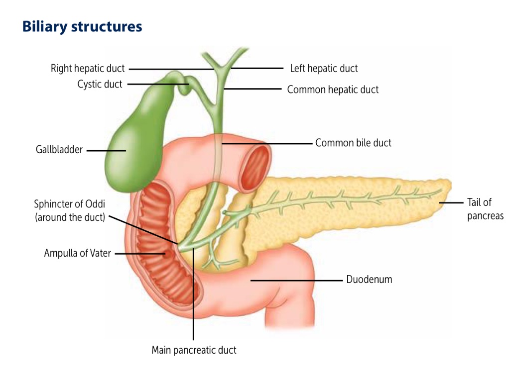
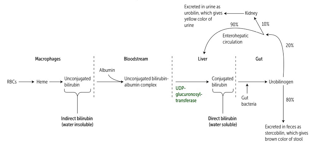
Cholelithiasis is obstruction of bile duct by a stone.

Obturation (obstruction, to close) of bile duct – it can be:

* Intrahepatic – blockage of intrahepatic bile ducts

* Extrahepatic – blockage of common bile duct (ductus choledochus).

Findings:

* malabsorption: bile salts do not enter the Small Intestine; no emulsification of fat.

*light coloured stool: due to lack of urobilin (which leads to lack of stercobilin).

*Jaundice (posthepatic, mechanic, obstructive): increased conjugated Bilirubin.

* Steatorrhea

The findings are specific for obstruction of bile duct and bile acid deficiency.

Indirect; Hemolytic; Prehepatic

Mixed; Parenchymal; Hepatic

Direct; Obstructive; Mechanic; Posthepatic

Stercobilin (faeces)

        ↑↑↑

Decreases (pale faces)

Absent (clay coloured faeces)

Type of bilirubin in blood

Unconjugated

Conjugated and Unconjugated

Conjugated

25.

A   48   year   old   patient   complained   about   intense   pain,   slight   swelling  and  reddening of  skin  over  the  joints,  temperature rise  up  to  38oC . Blood analysis revealed  high concentration of urates.  This condition might be caused by disturbed metabolism of:

Explanation

krushkrok No21 (2014)image

The end product of the purine nucleotides catabolism in humans and other primates is uric acid (urate) which is excreted in urine. Allopurinol and febuxostat inhibits Xanthine oxidase (XO). Hypoxanthine and Xanthine which is more soluble is excreted in urine. Purine nucleotides (adenine and guanine).  AMP – Adenosine monophosphate; GMP – Guanosine monophosphate

Pyrimidine (thymine, Uracil, cytosine); catabolism of thymine and uracil gives urea, while catabolism of cytosine gives β-alanine.

26.

Examination  of   a   patient  suffering  from  frequent  hemorrhages  in  the inner organs and mucous membranes revealed proline and lysine being included in collagen fibers. Impairment of their hydroxylation is caused by lack of the following vitamin:

Explanation

Vitamin C (ascorbic acid): found in fruits and vegetables; an antioxidant; also facilitates iron absorption by reducing it to Fe2+ state. It is necessary for hydroxylation of proline and lysine in collagen synthesis; necessary for dopamine β-hydroxylase, which converts dopamine to norepinephrine. Deficiency leads to: scurvy – swollen gums, bruising, petechiae, hemarthrosis, anemia, poor wound healing, perifollicular and subperiosteal hemorrhages, “corkscrew” hair; Weakened immune response.

Type III collagen is found in blood vessels; Type IV collagen is found in basement membrane. Deficiency in Vitamin C disrupts the second stage of collagen synthesis in fibroblasts (hydroxylation of collagen) which results in petechiae, bruising, hemarthrosis.

27. Untrained people  often  have  muscle pain after sprints as a result of lactate accumulation. This  might  be  caused  by intensification of the following biochemical process:

Explanation

Lactate, formed by the action of lactate dehydrogenase (converting pyruvate to lactate) is the final product of anaerobic glycolysis in eukaryotic cells. In organs or cells that are poorly vascularized and/or lack mitochondria, formation of lactate is the major fate of pyruvate as seen in lens, cornea of the eye, kidney medulla, testes, leukocytes and red blood cells.

Athletes that are exercising intensely for the short periods of time, such as in a sprint race, build up large amounts of lactate in their muscles as the result of anaerobic glycolysis. The “warming down” period of continual movement under aerobic conditions performed by athletes for approximately 15mins after a race increases circulation and removes lactate from the muscles.

28. Examination of a patient revealed typical presentations of collagenosis.  This pathology is characterized by increase  of the following urine index:

Explanation

Collagen, most abundant protein in human body; organizes and strengthens extracellular matrix. Collagen contains Gly-X-Y (X and Y are proline or lysine). Glycine(Gly) makes 1/3 of collagen. Oxyproline (hydroxyproline) is a major collagen amino acid which enables it to be regarded as a marker that reflects the catabolism of collagen.

29.

Labelled amino acids alanine  and tryptophane  were  injected   to  a  mouse in order  to  study  localization  of protein synthesis  in  its  cells.  The  labelled   amino acids will be accumulated near the following organellas:


Explanation

A large number of components are required for translation (synthesis of a protein). These include all the amino acids that are found in the finished product, the mRNA to be translated, tRNA, functional ribosomes, energy sources and enzymes as well as protein factors needed for initiation, elongation and termination of the polypeptide chain. In eukaryotic cells, the ribosomes are either “free” in the cytosol or are in close association with the endoplasmic reticulum (which is then known as the “rough” endoplasmic reticulum or RER). The RER – associated ribosomes are responsible for synthesizing proteins that are to be exported from the cell as well as those that are destined to become integrated into plasma, endoplasmic reticulum or golgi membranes or incorporated into lysosomes.

30.

A full-term newborn  child has yellowish skin and mucous  membranes. This might be probably caused by temporary deficiency of the following enzyme:

Explanation

image

In the hepatocyte, the solubility of unconjugated bilirubin is increased (i.e. it is made soluble) by the addition of two molecules of glucuronic acid to produce conjugated bilirubin. This process is reffered to as conjugation. This reaction is catalyzed by Uridine diphosphate (UDP) glucuronyltransferase – UGT. Varying degrees of deficiency of this enzyme result in Crigler-Najjar I & II and Gilbert syndrome; with Crigler-Najjar I being the most severe deficiency.

Deficiency of UGT inhibits conjugation and therefore increase unconjugated bilirubin in serum (Jaundice).

31.

A patient has low rate  of magnesium ions that  are  necessary  for affixion of ribosomes   to  the  endoplasmic  reticulum. It  is  known   that   it  causes  disturbance of protein biosynthesis.  At  what  stage  is protein biosynthesis  impaired?


Explanation

A large number of components are required for translation (synthesis of a protein). These include all the amino acids that are found in the finished product, the mRNA to be translated, tRNA, functional ribosomes, energy sources and enzymes as well as protein factors needed for initiation, elongation and termination of the polypeptide chain. In eukaryotic cells, the ribosomes are either “free” in the cytosol or are in close association with the endoplasmic reticulum (which is then known as the “rough” endoplasmic reticulum or RER). The RER – associated ribosomes are responsible for synthesizing proteins that are to be exported from the cell as well as those that are destined to become integrated into plasma, endoplasmic reticulum or golgi membranes or incorporated into lysosomes.

32.

Blood of a 12 year old boy presents low concentration of uric acid and accumulation  of  xanthine  and  hypoxanthine.  This child has  genetic  defect  of the  following enzyme:

Explanation

image

The end product of the purine nucleotides catabolism in humans and other primates is uric acid (urate) which is excreted in urine. Allopurinol and febuxostat inhibits Xanthine oxidase (XO). Hypoxanthine and Xanthine which is more soluble is excreted in urine. Purine nucleotides (adenine and guanine).  AMP – Adenosine monophosphate; GMP – Guanosine monophosphate

The last two steps in uric acid biosynthesis is catalyzed by xanthine oxidase.  Deficiency of this enzyme can lead to reduction in the uric acid concentration.

33. Examination of a patient suffering from cancer  of  urinary  bladder revealed  high rate  of serotonin and  hydroxyanthranilic acid. It is caused by excess of the following amino acid in the organism:

Explanation

Serotonin, also called 5-hydroxytryptamine, is synthesized and stored at several sites in the body. It can be found in the intestinal mucosa, central nervous system and in platelets. Serotonin is synthesized from tryptophan, which is hydroxylated. The product, 5-hydroxytryptophan is decarboxylated to serotonin, which is also degraded by monoamine oxidase (MAO). Serotonin has multiple physiologic roles, including pain perception, affective disorders, and regulation of sleep, temperature and blood pressure.

3-hydroxyanthranilic acid is an intermediate in the metabolism of tryptophan.

34.

Study of conversion  of a food colouring agent  revealed  that  neutralization of this  xenobiotic   takes  place  only  in  one phase  - microsomal   oxidation.   Name  a component of this phase:

Explanation

     The P450 system is impotant for the metabolism of many endogenous compounds (such as steroids, lipids etc) and for the biotransformation of exogenous substances (xenobiotics). Cytochrome P450, designated as CYP, is a superfamily of heme-containing isozymes that are located in most cells but are primarily found in the liver and GIT. Phase I reactions utilizing the P450 system (also called microsomal mixed function oxidases). The oxidation proceeds by the xenobiotic binding to the oxidized form of cytochrome P450 and then O2 is introduced through a reductive step, coupled to NADPH: cytochrome P450 oxidoreductase.

35.

A child has mental  and physical retardation, grave damage  of internal connective tissue.  Urine  analysis  reveals keratan sulfates.  What  metabolic process is disturbed?

Explanation

The Mucopolysaccharidoses are hereditary disorders that are clinically progressive. They are characterized by accumulation of glycosaminoglycans in various tissues, causing varied symptoms such as skeletal and extracellular matrix deformities and mental retardation. Mucopolysaccharidoses are caused by a deficiency of any one of the lysosomal hydrolases normally involved in the degradation of heparin sulfate and/or dermatan sulfate. This results in the presence of oligosaccharides in the urine because of incomplete lysosomal degradation of glycosaminoglycans.

36.

A newborn child was found  to have reduced intensity of sucking, frequent vomiting, hypotonia. Urine and blood exhibit increased  concentration of citrulline. What metabolic process is disturbed?

Explanation

image

In urea or ornithine cycle, the first two reactions leading to the synthesis of urea occur in the mitochondria, whereas the remaining cycle enzymes are located in the cytosol. The first reaction is formation of carbomoyl phosphate and the second reaction is formation of citrulline. Ornithine and citrulline are basic amino acids that participate in the urea cycle. The release of the high energy phosphate of carbomoyl phosphate as inorganic phosphate drives the reaction in the forward direction. The reaction product – Citrulline is transported to the cytosol where it condenses with aspartate to form argininosucinate. Increased concentration of citrulline in urine and blood indicates a defect in ornithine or urea cycle.

37. On  an  electron   micrograph  a  scientist  has  identified   a  structure  formed by  eight  histone  proteins and  a  part  of DNA molecule which makes about 1,75 revolutions around the molecules.  Which structure has been identified?

Explanation

image    

Genes are contained in a complex of DNA and proteins (mostly histones) called chromatin and its basic unit of structure is the nucleosome. Each nucleosome is composed of an octamer (8) of histone proteins and approximately 140 base pairs of DNA. Nucleosomes themselves are joined into clusters by binding of DNA existing between nucleosomes (linker DNA) with other histone proteins. Nucleosomes keep the DNA tightly coiled such that it cannot be transcribed.

38.

A    newborn   develops    dyspepsia after   the   milk  feeding.   When   the   milk is substituted by the  glucose  solution the  dyspepsia  symptoms  disappear. The newborn  has the subnormal activity of the following enzyme:

Explanation

    Lactase (β-galactosidase) cleaves lactose (in milk) producing galactose and glucose. Hereditary deficiencies of lactase have been reported in infants and children with dissacharide intolerance. Treatment for this disorder is to reduce consumption of milk. This is seen when the milk is substituted by glucose solution.

39. Some  students  developed myodynia after  continuous physical  activity  during physical  education. The  reason  for  such condition was accumulation of lactic acid in the  skeletal  muscles.  It was generated in the students’ bodies  after  activation of the following process:

Explanation

Lactate, formed by the action of lactate dehydrogenase (converting pyruvate to lactate) is the final product of anaerobic glycolysis in eukaryotic cells. In organs or cells that are poorly vascularized and/or lack mitochondria, formation of lactate is the major fate of pyruvate as seen in lens, cornea of the eye, kidney medulla, testes, leukocytes and red blood cells.

Aerobic glycolysis progresses to citric acid cycle from pyruvate. The cycle occurs totally in the mitochondria.

       Athletes that are exercising intensely for the short periods of time, such as in a sprint race, build up large amounts of lactate in their muscles as the result of anaerobic glycolysis. The “warming down” period of continual movement under aerobic conditions performed by athletes for approximately 15mins after a race increases circulation and removes lactate from the muscles.

40.

During  starvation muscle proteins break up into free amino acids. These compounds will be the most probably involved into the following process:

Explanation

     Gluconeogenesis is making of glucose from non-carbohydrate sources i.e. fat and proteins. During a prolonged fast, hepatic glycogen stores are depleted and glucose is formed from precursors such as lactate, pyruvate, glycerol (derived from backbone of triacylglycerols) and α-ketoacids (derived from the catabolism of glucogenic amino acids). Amino acids derived from hydrolysis of tissue proteins (e.g. muscle proteins) are the major sources of glucose during a fast. α-ketoacids such as oxaloacetate and α-ketoglutarate are derived from the metabolism of glucogenic amino acids. Amino acids whose catabolism yields pyruvate or one of the intermediates of citric acid cycle (CAC) are termed glucogenic or glycogenic e.g. alanine, arginine, aspartate, histidine etc.

41. A 1-year-old baby has been hospitalised  for  body  and  limbs  lesions.  Examination  revealed  carnitine deficiency in the child’s muscles. A biochemical  reason  for this pathology is the disorder  of:

Explanation

image

The major pathway for catabolism of saturated fatty acids is a mitochondrial pathway called β-oxidation. After a long-chain fatty acid (LCFA) enters a cell, it is converted in the cytosol to its Co-A derivative. Because β-oxidation occurs in the mitochondrial matrix, the fatty acid must be transported across inner mitochondrial membrane which is impermeable to Co-A. therefore, a specialized carrier transports the long chain acyl group from the cytosol into the mitochondrial matrix. This carrier is carnitine and this rate-limiting transport process is called the carnitine shuttle. Since carnitine helps the mitochondria utilize energy, it plays a critical role in reducing the occurrence and impact of obesity. In addition to helping the mitochondria burn fat as energy, carnitine is also vital for removing waste products from mitochondria. Obesity and aging contribute to low carnitine levels, which compromises mitochondrial performance and increases insulin resistance, promoting further obesity and carnitine reduction.

42. It  was  found  out  that  some compounds, for instance  fungi toxins and some  antibiotics   can  inhibit   activity  of RNA-polymerase.  What  process  will be disturbed in a cell in case of inhibition  of this enzyme?

Explanation

     The process of transcription of a typical gene can be divided into 3 phases: initiation, elongation and termination. The process of transcription begins with the binding of the RNA polymerase holoenzyme to a regionof the DNA known as the promoter. RNA polymerase is a multisubunit enzyme that recognizes a nucleotide sequence (the promoter region) at the beginning of a length of DNA that is to be transcribed. It makes a complementary RNA copy of the DNA template strand and then recognizes the end of the DNA sequence to be transcribed (the termination region).

43. A patient complained about  dizziness, memory  impairment, periodical convulsions. It was revealed that these changes were caused by a product of decarboxylation of  glutamic  acid.  Name this product:

Explanation

Certain amino acids undergo decarboxylation that means the removal of their α-carboxyl group resulting in liberation of CO2 and formation of biogenic amines. Biogenic amines are physiologically active substances such as hormones, neurotransmitters etc. decarboxylation of amino acids:

image

·        Tryptophan → Niacin → NAD+/NADP+

         Tryptophan → Serotonin →Melatonin

·        Histidine → Histamine

·        Glutamine → GABA

         Glutamine → Glutathione

44.

During  hypersensitivity test a patient got subcutaneous injection  of an antigen which  caused  reddening of skin,  edema, pain  as a result  of histamine action.  This biogenic amine  is generated as a result of transformation of the  following  histidine amino acid:

Explanation

     Certain amino acids undergo decarboxylation that means the removal of their α-carboxyl group resulting in liberation of CO2 and formation of biogenic amines. Biogenic amines are physiologically active substances such as hormones, neurotransmitters etc. decarboxylation of amino acids:

image

·        Tryptophan → Niacin → NAD+/NADP+

         Tryptophan → Serotonin →Melatonin

·        Histidine → Histamine

·        Glutamine → GABA

         Glutamine → Glutathione

45.

Vitamin B1  deficiency causes disturbance   of   oxidative    decarboxylation  of  α-ketoglutaric acid.  This  leads  to the  impaired synthesis  of  the  following coenzyme:

Explanation

     Thiamine (vitamin B1): thiamine pyrophosphate (TPP) is the biologically active form of the vitamin, formed by the transfer of a pyrophosphate group from ATP to thiamine. Biological role of TPP: it is a component of pyruvate dehydrogenase and α-ketoglutarate dehydrogenase complexes catalyzing the reactions of oxidative decarboxylation of pyruvate and α-ketoglutarate (kreb’s cycle) i.e. it promotes energy formation from carbohydrates and lipids. It’s also a component of transketolase (pentose phosphate pathway of glucose oxidation) essential for fats and nucleic acids synthesis.

46.

Jaundice treatment involves administration of barbiturates inducing the synthesis  of UDP-glucuronyl transferase. A medicinal  effect is caused by the production of:

Explanation

Crigler Najjar syndrome type II is a rare hereditary disorder and is due to a less severe defect in the bilirubin conjugation. It is believed that hepatic UDP-glucuronyltransferase that catalyzes the addition of second glucuronyl group is defective. It is treated with Phenobarbital ( a barbiturate), because it induces the action of UDP-glucuronyltransferase, thereby producing more conjugated (direct) bilirubin.

47.

It was revealed that  T-lymphocytes were  affected   by  HIV.   Virus  enzyme - reverse transcriptase   (RNA-dependent DNA-polymerase) - catalyzes the synthesis of:

Explanation

     Human immunodeficiency virus (HIV): diploid genome (2 molecules of RNA). The 3 structural genes(i.e. proteins coded for by the genes) are:

·        env (gp 120 and gp 41): formed from cleavage of gp 160 to form envelope glycoproteins. gp 120 is for attachment to host CD4+ T cell. gp 41 is for fusion and entry.

·        Gag (p24): capsid protein

·        pol: reverse transcriptase, aspartate protease, integrase.

ELISA/Western blot (immunoblot) tests look for antibodies to the viral proteins listed above.

Reverse transcriptase synthesizes dsDNA (ds-double stranded) from genomic RNA (mRNA); dsDNA integrates into host genome. Virus binds CD4 as well as a coreceptor, either CCR5 on macrophages (early infection) or CXCR4 on I cells (late infection).

·        Homozygous CCR5 mutation – immunity

·        Heterozygous CCR5 mutation – slower course.

48. A  patient has  an  increased pyruvate concentration  in  blood,   most   of   it  is excreted with the urine. What kind of avitaminosis  has this patient?

Explanation

     Thiamine (vitamin B1): thiamine pyrophosphate (TPP) is the biologically active form of the vitamin, formed by the transfer of a pyrophosphate group from ATP to thiamine. Biological role of TPP: it is a component of pyruvate dehydrogenase and α-ketoglutarate dehydrogenase complexes catalyzing the reactions of oxidative decarboxylation of pyruvate and α-ketoglutarate (kreb’s cycle) i.e. it promotes energy formation from carbohydrates and lipids. It’s also a component of transketolase (pentose phosphate pathway of glucose oxidation) essential for fats and nucleic acids synthesis.

Pyruvate to acetyl CoA + CO2 reaction

            If pyruvate dehydrogenase cannot function properly due to vitamin B1 deficiency, then pyruvate will be accumulated in blood because it can’t be broken down.

49.

Urine  analysis  of  a  12-year-old  boy reveals high concentration of all aliphatic amino  acids  with  the  highest  excretion of cystine and cysteine. US of kidneys revealed  kidney concrements. What is the most likely pathology?

Explanation

     Cystinuria is a disorder of the proximal tubules reabsorption of filtered cystine and dibasic amino acids (lysine, ornithine, arginine). The inability to reabsorb cystine leads to accumulation and subsequent precipitation of stones of cystine in the urinary tract. It is an inherited disorder of amino acid transport. The disease expresses itself clinically by the precipitation of cystine to form kidney stones (calculi), which can block the urinary tract. Oral hydration is an important part of treatment for this disorder. Cystine is formed from two molecules of cysteine joined together.

50. The greater amount of nitrogen is excreted from  the  organism  in  form  of urea. Inhibition of urea synthesis and accumulation of  ammonia in  blood  and tissues are induced by the decreased activity of the following liver enzyme:

Explanation

image     

Urea is the major disposal form of amino groups derived from amino acids and account for about 90% of the nitrogen-containing components of urine. Formation of carbomoyl phosphate by carbomoyl phosphate synthetase I is driven by cleavage of two molecules of ATP. Ammonia is incorporated into carbamoyl phosphate. Ultimately, the nitrogen atom derived from this ammonia becomes one of the nitrogens of urea. NB: carbamoyl phosphate synthetase II participates in the biosynthesis of pyrimidines. However, when the function of carbomoyl phosphate synthetase I is compromised, blood ammonia levels rise and urea synthesis is disturbed.

51.

A  doctor  recommends a  patient with  duodenal ulcer  to  drink  cabbage  and potato  juice   after   the   therapy  course. Which substances  contained in these vegetables  help  to  heal  and  prevent the ulcers?

Explanation

     Cabbage juice is one of the most healing nutrients for ulcer repair as it is a huge source of vitamin U (vitamin U is actually not a vitamin but an enzyme known as S-methylmethionine). Research shows that vitamin U, administered as raw cabbage juice, is effective in promoting the rapid healing of peptic ulcers. Potatoes on the other hand are rich sources of vitamins and minerals. Several studies, such as the one published in the June 2008 edition of the “Journal of vascular nursing”, have shown reduced levels of vitamin A in patients suffering from leg ulcers, indicating a connection between the two, probably because vitamin A helps to form and maintain healthy skin, mucous membrane and teeth. Vitamin A and the amino acid glutamine help to regenerate healthy epithelial cells.

52. A   46-year-old   patient  consulted   a doctor complaining about joint pain that becomes   stronger  the   day   before   the weather   changes.    Blood    examination revealed an increased concentration of uric acid. The most probable cause of the disease is the intensified disintegration of the following substance:

Explanation

image

The end product of the purine nucleotides catabolism in humans and other primates is uric acid (urate) which is excreted in urine. Allopurinol and febuxostat inhibits Xanthine oxidase (XO). Hypoxanthine and Xanthine which is more soluble is excreted in urine. Purine nucleotides (adenine and guanine).  AMP – Adenosine monophosphate; GMP – Guanosine monophosphate

Pyrimidine (thymine, Uracil, cytosine); catabolism of thymine and uracil gives urea, while catabolism of cytosine gives β-alanine.

53.

A  patient has  pellagra.  Interrogation revealed  that he had lived mostly on maize for a long time  and  eaten  little  meat. This disease had been caused by the deficit of the following substance  in the maize:

Explanation

Vit. B3 (Niacin, PP): coenzyme forms NAD+, NADP+ is derived from tryptophan. Synthesis requires Vit. B2 and B6.

Vitamin B3 (niacin) is very high in fish and meat products.

54.

Researchers   isolated    5   isoenzymic forms  of lactate  dehydrogenase from  the human blood serum and studied their properties. What  property indicates  that the  isoenzymic  forms  were  isolated  from the same enzyme?

Explanation

     Isoenzyme is any of several forms of an enzyme that all catalyze the same reaction but may differ in reaction rate, inhibition by various substances, electrophoretic mobility or immunologic properties. Examples of important enzymes with isoenzymic forms are:

·        Lactate dehydrogenase: LDH1, LDH2, LDH3, LDH4, LDH5, (5 isoenzymic forms).

·        Creatine kinase (CK): CK-MM; CK-MB; CK-BB (3 isoenzymic forms)

55.

A  10-year-old   girl  has  a  history   of repeated acute respiratory viral infection. After  recovering  she presents with multiple petechial hemorrhages on the sites of friction  from  clothing  rubbing   the  skin. What kind of hypovitaminosis has this girl?

Explanation

Vitamin C (ascorbic acid): found in fruits and vegetables; an antioxidant; also facilitates iron absorption by reducing it to Fe2+ state. It is necessary for hydroxylation of proline and lysine in collagen synthesis; necessary for dopamine β-hydroxylase, which converts dopamine to norepinephrine. Deficiency leads to: scurvy – swollen gums, bruising, petechiae, hemarthrosis, anemia, poor wound healing, perifollicular and subperiosteal hemorrhages, “corkscrew” hair; Weakened immune response.

Type III collagen is found in blood vessels; Type IV collagen is found in basement membrane. Deficiency in Vitamin C disrupts the second stage of collagen synthesis in fibroblasts (hydroxylation of collagen) which results in petechiae, bruising, hemarthrosis.

56.

A  patient  presents with  dysfunction of cerebral cortex accompanied by epileptic  seizures.  He  has  been  administered  a biogenic amine  synthesized from glutamate and responsible for central inhibition.  What substance  is it?

Explanation

Certain amino acids undergo decarboxylation that means the removal of their α-carboxyl group resulting in liberation of CO2 and formation of biogenic amines. Biogenic amines are physiologically active substances such as hormones, neurotransmitters etc. decarboxylation of amino acids:

image

·        Tryptophan → Niacin → NAD+/NADP+

         Tryptophan → Serotonin →Melatonin

·        Histidine → Histamine

·        Glutamine → GABA

         Glutamine → Glutathione

↑glutamate → ↑GABA (inhibitory neurotransmitter)
57.

Osteolaterism is characterized by a decrease in  collagen  strength   caused  by much  less  intensive  formation  of  cross-links in collagen fibrils. This phenomenon is caused by the low activity of the following enzyme:

Explanation

Osteolaterism results in conditions of deficiency or non-functional lysyl oxidase. Lysyl oxidase is involved in the cross-linking of elastin and collagen resulting in disorganized connective tissue formation. Lysyl oxidase is an extracellular copper enzyme that catalyzes formation of aldehydes from lysine residues in collagen and elastin precursors. This results in cross-linking collagen and elastin, which is essential for stabilization of collagen fibrils and for the integrity and elasticity of mature elastin.

58.

A  patient has  been  diagnosed  with alkaptonuria. Choose  an  enzyme  whose deficiency can be the reason for this pathology:

Explanation

image

Ochronosis (Alkaptonuria): congenital deficiency of homogentisate oxidase (homogentisic acid oxidase) in the degradative pathway of tyrosine to Fumarate → pigment-forming homogentisic acid accumulates (homogentisuria) in tissues. Autosomal recessive. Usually benign. Urine turns black on prolonged exposure to air. May have debilitating arthralgias (homogentisic acid toxic to cartilage).

59. Emotional stress  causes  activation of hormone-sensitive triglyceride  lipase in the adipocytes. What secondary mediator takes part in this process?

Explanation

The mobilization of stored fat requires the hydrolytic release of fatty acids and glycerol from their triacylglycerol (TAG) form. This process is initiated by hormone-sensitive lipase, which removes a fatty acid from carbon 1 and/or carbon 3 of the TAG. This enzyme is activated when phosphorylated by cyclic AMP (cAMP)-dependent protein kinase. cAMP is produced in the adipocyte when one of several hormones (such as epinephrine or glucagon – stress hormones) binds to receptors on the cell membrane and activates adenylyl cyclase.

60.

A 36 year old female patient has a history of collagen disease. Urine analysis is likely to reveal an increased concentration of the following metabolite:

Explanation

Collagen, most abundant protein in human body; organizes and strengthens extracellular matrix. Collagen contains Gly-X-Y (X and Y are proline or lysine). Glycine(Gly) makes 1/3 of collagen. Oxyproline (hydroxyproline) is a major collagen amino acid which enables it to be regarded as a marker that reflects the catabolism of collagen.

61.

Vitamin  A together with specific cytoreceptors penetrates through the nuclear   membranes, induces  transcription  processes  that  stimulate growth  and differentiation of cells. This biological function  is realized  by the following form of vitamin A:

Explanation

Vitamin A and retinoids play an important role in the orderly differentiation of mucus-secreting epithelium. All-trans-retinoic acid, a potent acid derivative of vitamin A, has the highest affinity for retinoic acid receptors (RARs) compared with other retinoids. Activation of RARs by their ligands causes the release of corepressor and the obligatory formation of heterodimers with another retinoid receptor, known as the retinoic X receptor (RXR). The RAR/RXR heterodimers bind to retinoic acid response elements located in the regulatory regions of genes that encode receptors for growth factors, tumor suppressor genes and secreted proteins. Through these effects, they regulate cell growth and differentiation, cell cycle control and other biologic responses.
62.

After severe viral hepatitis a 4 year old boy presents with vomiting,  occasional loss of consciousness, convulsions. Blood test revealed hyperammoniemia. Such condition is caused by a disorder of the following biochemical hepatic process:

Explanation

image     

Hyperammonemia can be acquired (e.g. liver disease) or hereditary (e.g. urea cycle enzyme deficiencies). Acquired hyperammonemia may be a result of an acute process e.g. viral hepatitis, ischemia or hepatotoxins. As a result, the detoxification of ammonia (i.e. its conversion to urea) is severely impaired, leading to elevated levels of circulating ammonia.

63.

Before the cells can utilize the glucose, it is first transported from the extracellular space  through the  plasmatic   membrane inside them. This process is stimulated by the following hormone:

Explanation

In the liver and muscle, insulin increases glycogen synthesis. In the muscle and adipose tissue, insulin increases glucose uptake by increasing the number of glucose transporters (GLUT-2) in the cell membrane. Insulin is an anabolic hormone; it increases glucose uptake, glycogen synthesis, protein synthesis, fat synthesis; and decrease gluconeogenesis, glycogenolysis, lipolysis. Gluconeogenesis is the production of glucose from non-carbohydrate sources.

Glucagon is a functional antagonist of insulin.

64.

A   2 year   old   child   with   mental and physical retardation has been delivered to a hospital. He presents with frequent vomiting   after   having   meals. There is phenylpyruvic acid in urine. Which metabolism abnormality is the  reason for this pathology?

Explanation

     Phenylketones – phenylacetate, phenyllactate and phenylpyruvate. These compounds are not normally produced in significant amounts in the presence of functional phenylalanine hydroxylase, but are elevated in phenylketonuria. These metabolites give urine a characteristic musty (“mousey”) odour. The disease acquired its name from the presence of a phenylketone (now known to be phenylpyruvate) in the urine.

image

Phenylketonuria (PKU) is caused by a deficiency of phenylalanine hydroxylase. PKU is the most common clinically encountered inborn error of amino-acid metabolism. Biochemically, it is characterized by accumulation of phenylalanine and a deficiency of tyrosine. It may also be caused by deficiencies in tetrahydrobiopterin cofactor (BH4) or in dihydropteridine (BH2) reductase, which regenerates BH4 from BH2.

65.

Examination  of  a  child  who  hasn’t got fresh  fruit  and  vegetables  during  winter revealed numerous subcutaneous hemorrhages, gingivitis, carious cavities in teeth.  What vitamin combination should be prescribed in this case?

Explanation

Vitamin C (ascorbic acid): found in fruits and vegetables; an antioxidant; also facilitates iron absorption by reducing it to Fe2+ state. It is necessary for hydroxylation of proline and lysine in collagen synthesis; necessary for dopamine β-hydroxylase, which converts dopamine to norepinephrine. Deficiency leads to: scurvy – swollen gums, bruising, petechiae, hemarthrosis, anemia, poor wound healing, perifollicular and subperiosteal hemorrhages, “corkscrew” hair; Weakened immune response.

Type III collagen is found in blood vessels; Type IV collagen is found in basement membrane. Deficiency in Vitamin C disrupts the second stage of collagen synthesis in fibroblasts (hydroxylation of collagen) which results in petechiae, bruising, hemarthrosis.

Vitamin P (rutin) – permeability vitamin. They reduce permeability of blood vessels, especially capillaries. It prevents hyaluronic acid (the basic compound of the extracellular matrix) from degeneration by inhibition of the enzyme hyaluronidase. Ascorutin is a drug containing vitamin C and P; it is used to decrease the permeability of blood vessels.

66.

An experimental animal that was kept on  protein-free  diet  developed  fatty  liver  infiltration,   in  particular as  a  result of deficiency  of methylating agents.  This is caused by disturbed generation of the following metabolite:

Explanation

Hepatic steatosis can occur when humans are deprived of choline.

Choline + Phosphatidic acid → Phosphatidylcholine (lecithin, PC). In the liver PC can also be synthesized from phosphatidylserine (PS) and phosphatidylethanolamine (PE), when free choline levels are low, because it exports significant amounts of PC in bile and as a component of serum lipoproteins (needed for fat metabolism)

PS → PE →→→ PC. 3 methylation reactions between PE and PC. S-adenosylmethionine is the methyl group donor. If choline, phosphatidylcholine or methionine is deficient, there will be abnormal phospholipid synthesis, oxidative damage caused by mitochondrial dysfunction, lipoprotein secretion (remember, if VLDL cannot be secreted it will be accumulated & cause fatty liver degeneration as seen in hepatic steatosis). PC is also a major lipid component of lung surfactant.

Vitamin B12 is a cofactor for homocysteine methyltransferase and methylmalonyl-CoA mutase. Both vitamin B12 and B6 are needed in fatty acid (FA) metabolism.

67.

Characteristic   sign   of   glycogenosis is  muscle   pain   during   physical   work. Blood examination reveals usually hypoglycemia. This  pathology  is caused by congenital deficiency  of the  following enzyme:

Explanation

     Glycogen phosphorylase is the rate-determining enzyme in Glycogenolysis (break down of glycogen). Glycogen phosphorylase cleaves glycogen to glucose 1-phosphate (first step in glycogenolysis). This pathway mobilizes stored glycogen in liver to replenish used glucose. It also breaks down glycogen in muscle to glucose, to produce energy during physical work. If this enzyme is deficient, then used glucose cannot be replaced leading to hypoglycemia.

In lysosomal glycosidase, there is still normal blood sugar levels (no hypoglycemia). α-amylase and ɣ-amylase is involved in digestion of polysaccharides. Glucose 6-phosphate dehydrogenase (G6PD) is the rate-limiting enzyme in pentose phosphate pathway which catalyzes an irreversible oxidation of glucose 6-phosphate to 6-phosphogluconolactone.

68.

At  the  stage  of  translation  in  the rough  endoplasmic reticulum,  the ribosome  moves along the mRNA. Amino acids are joined together by peptide bonds in a specific sequence, and thus polypeptide synthesis takes place. The sequence of amino acids in a polypeptide corresponds to the sequence  of:

Explanation

The pathway of protein synthesis translates the 3-letter alphabet of nucleotide sequences on mRNA into the twenty-letter alphabet of amino acids that constitute proteins. mRNA convey genetic information from DNA to the ribosome, where they specify the amino acid sequence of the protein products of gene expression. As in DNA, mRNA genetic information is in the sequence of nucleotides, which are arranged into codons consisting of 3 bases each. Each codon encodes for a specific amino acid, except the stop codons, which terminate protein synthesis. The mRNA is translated from its 5’-end to its 3’-end, producing a protein synthesized from its amino-terminal end to its carboxyl-terminal end.

69. Children with Lesch-Nyhan syndrome have a severe form of hyperuricemia  accompanied by the  formation of tophi,  urate  calculi  in the  urinary  tracts, as well as serious neuro-psychiatric disorders.  The  cause  of this  disease  is the reduced activity of the following enzyme:

Explanation

image

Lesch-Nyhan syndrome: defective purine salvage due to absent hypoxanthine-guanine phosphoribosyl transferase (HGPRT), which converts hypoxanthine to inosine monophosphate (IMP) and guanine to Guanosine monophosphate (GMP). Results in excess uric acid production and de novo purine synthesis. This syndrome is X-linked recessive. Findings: intellectual disability, self-mutilation, aggression, hyperuricemia, gout, dystonia. It can be treated with allopurinol and febuxostat.

70.

The genetic defect of pyruvate carboxylase deficiency is the cause of delayed physical and mental development and  early  death  in children.  This  defect is characterized by lacticemia,  lactaciduria,  disorder of a number  of metabolic pathways.  In particular, the following process is inhibited:

Explanation

In mitochondria, pyruvate carboxylase catalyzes the conversion of pyruvate to oxaloacetate (in gluconeogenesis). This enzyme requires biotin and ATP. Oxaloacetate can replenish citric acid cycle (CAC) or be used in gluconeogenesis. So, a deficiency of pyruvate decarboxylase impairs both CAC and gluconeogenesis.

71.

An experiment proved  that UV- irradiated skin cells of patients with xeroderma pigmentosum restore the native structure of DNA  slower than the cells of  healthy   people   due  to  the  defect  in repair  enzyme.  What  enzyme  takes  part in this process?

Explanation

Exposure of a cell to ultraviolet (UV) light can result in the covalent joining of two adjacent pyrimidines (usually thymine), producing a dimer. These thymine dimers prevent DNA polymerase from replicating the DNA strand beyond the site of dimer formation. First, a UV-specific endonuclease recognizes the dimer an cleaves the damaged strand on both the 5’-side and 3’-side of the dimer. Pyrimidine dimers can be formed in the skin cells of humans exposed to unfiltered sunlight. In the rare genetic disease Xeroderma pigmentosum, the cells cannot repair the damaged DNA because UV-specific endonuclease is defective, resulting in extensive accumulation of mutations and consequently, skin cancers.

72. One  of the factors that  cause obesity is the  inhibition  of  fatty  acids  oxidation due to:

Explanation

image

The major pathway for catabolism of saturated fatty acids is a mitochondrial pathway called β-oxidation. After a long-chain fatty acid (LCFA) enters a cell, it is converted in the cytosol to its Co-A derivative. Because β-oxidation occurs in the mitochondrial matrix, the fatty acid must be transported across inner mitochondrial membrane which is impermeable to Co-A. therefore, a specialized carrier transports the long chain acyl group from the cytosol into the mitochondrial matrix. This carrier is carnitine and this rate-limiting transport process is called the carnitine shuttle. Since carnitine helps the mitochondria utilize energy, it plays a critical role in reducing the occurrence and impact of obesity. In addition to helping the mitochondria burn fat as energy, carnitine is also vital for removing waste products from mitochondria. Obesity and aging contribute to low carnitine levels, which compromises mitochondrial performance and increases insulin resistance, promoting further obesity and carnitine reduction.

73.

For   the   study   of  serum   proteins various physical and physicochemical methods  can be used. In particular, serum albumins  and  globulins  can be separated by this method:


Explanation

Electrophoresis uses the principle of electric charge size and shape to separate substances. Proteins carry a positive or a negative electrical charge and they move in fluid when placed in an electrical field. The two major types of protein present in the serum are albumin and globulin proteins. Albumin is the major protein component of serum and represents the largest peak that lies closest to the positive electrode. Globulins comprise a much smaller fraction of the total serum protein but represent the primary focus of interpretation of serum protein electrophoresis. Five (5) globulin categories are expressed – α1, α2, (alpha 1 & 2), β1, β2 (beta 1 & 2) and ɣ (gamma). ɣ is the closest to the positive electrode.

74. Enzymatic  jaundices  are accompanied  by  abnormal activity  of  UDP-glucuronyl   transferase. What  compound is accumulated in blood  serum  in case of these pathologies?

Explanation

image

In the hepatocyte, the solubility of unconjugated bilirubin is increased (i.e. it is made soluble) by the addition of two molecules of glucuronic acid to produce conjugated bilirubin. This process is reffered to as conjugation. This reaction is catalyzed by Uridine diphosphate (UDP) glucuronyltransferase – UGT. Varying degrees of deficiency of this enzyme result in Crigler-Najjar I & II and Gilbert syndrome; with Crigler-Najjar I being the most severe deficiency.

Deficiency of UGT inhibits conjugation and therefore increase unconjugated bilirubin in serum (Jaundice).

75.

It   is  known   that   the   monoamine oxidase (MAO) enzyme plays an important part in the metabolism of catecholamine neurotransmitters. In what way does the enzyme inactivate  these neurotransmitters (norepinephrine, epinephrine, dopamine)?

Explanation

image

Norepinephrine (noradrenaline), epinephrine (adrenaline) and dopamine are catecholamines produced in chromaffin cells of adrenal medulla from tyrosine. The catecholamines are inactivated by oxidative deamination catalyzed by monoamine oxidase (MAO) and by O-methylation carried out by Catechol-O-methyltransferase. The metabolic products of these reactions are excreted in the urine as vanillymandelic acid from epinephrine and norepinephrine; and homovanillic acid from dopamine.

76. A  20-year-old  male  patient complains  of  general   weakness,   rapid   fatigability, irritability, decreased performance, bleeding   gums,  petechiae  on  the   skin. What  vitamin  deficiency  may be a cause of these changes?

Explanation

Vitamin C (ascorbic acid): found in fruits and vegetables; an antioxidant; also facilitates iron absorption by reducing it to Fe2+ state. It is necessary for hydroxylation of proline and lysine in collagen synthesis; necessary for dopamine β-hydroxylase, which converts dopamine to norepinephrine. Deficiency leads to: scurvy – swollen gums, bruising, petechiae, hemarthrosis, anemia, poor wound healing, perifollicular and subperiosteal hemorrhages, “corkscrew” hair; Weakened immune response.

Type III collagen is found in blood vessels; Type IV collagen is found in basement membrane. Deficiency in Vitamin C disrupts the second stage of collagen synthesis in fibroblasts (hydroxylation of collagen) which results in petechiae, bruising, hemarthrosis.

Vitamin B2 (riboflavin) deficiency – growth retardation, glossitis, conjunctivitis

Vitamin B1 (thiamine) deficiency – Beri-Beri (polyneuritis)

Vitamin A (retinol) deficiency – Night blindness

Vitamin B9 (folic acid) deficiency – macrocytic megaloblastic anemia

77.

By the decarboxylation of glutamate in the CNS an inhibitory  mediator is formed. Name it:

Explanation

Certain amino acids undergo decarboxylation that means the removal of their α-carboxyl group resulting in liberation of CO2 and formation of biogenic amines. Biogenic amines are physiologically active substances such as hormones, neurotransmitters etc. decarboxylation of amino acids:

image

·        Tryptophan → Niacin → NAD+/NADP+

         Tryptophan → Serotonin →Melatonin

·        Histidine → Histamine

·        Glutamine → GABA

         Glutamine → Glutathione

78.

An  unconscious  patient was taken  by ambulance  to  the  hospital.   On  objective  examination  the   patient  was  found to  have  no  reflexes,  periodical   convulsions, irregular breathing. After  laboratory examination the patient was diagnosed with   hepatic    coma.   Disorders  of   the central nervous system develop due to the accumulation of the following metabolite:

Explanation

Substances absorbed into the bloodstream from the intestine pass through the liver, where toxins are normally removed. Many of these toxins (such as ammonia) are normal breakdown products of the digestion of protein. In hepatic encephalopathy (hepatic coma), toxins are not removed because liver function is impaired. Ammonia is produced by amino acid metabolism and intestinal urease-positive bacteria. In physiological conditions, it is mostly present as ammonium (NH4+) in serum. The urea or ornithine cycle, which is fully expressed in the liver exclusively, serves to converts NH4+ to urea prior to renal excretion and to maintain low serum concentrations. In hepatic coma, when the liver cannot remove toxins and urea cycle is not functional, all this occurs:

NH3 + α-ketoglutarate → Glutamate

α-ketoglutarate is used up which leads to:

·        ↑glutamate → ↑GABA (inhibitory neurotransmitter)

·        Inhibition of citric acid cycle/tricarboxylic acid cycle; this causes impairment of ATP formation.

·        Inhibition of metabolism of amino acids (impairment of transamination reactions).


NH3 + Glutamate → Glutamine

Glutamine is an amide of glutamic acid which provides a non-toxic storage and transport form of ammonia (NH3). Ammonia increase synthesis of glutamine in brain. Accumulation of glutamine in brain results in elevation of osmotic pressure in nervous cells leading to brain edema.


                                    NH3 + H+ → NH4+

In blood ammonia (NH3) is represented as ammonium ion (NH4+). Accumulation of ammonium ion impairs transport of ions (Na+, K+) through cell membranes and failure of transmission of nerve impulse.

            Urea cycle takes place exclusively in the liver, so in hepatic coma, urea level is low. Glutamine toxicity in brain is dependent on increased ammonia concentration.

Bilirubin toxicity will most likely be related to increase hemolysis, which is not the case in this question. Histamine is a biogenic amine produced from the amino acid histidine.

79. A   number  of  diseases   can  be  diagnosed  by  evaluating activity  of  blood transaminases. What vitamin is one of cofactors  of these enzymes?

Explanation

Vitamin B6 (pyridoxine): coenzyme forms – Pyridoxal phosphate (PALP)

                                                                         Pyridoxamine Phosphate (PAMP)

Components of pyridoxal enzymes used in transamination (e.g. ALT and AST); decarboxylation reactions, glycogen phosphorylase, transformation of tryptophan to Niacin (Vit B3), active transport of amino acids through the cell membrane, synthesis of cystathionine, heme, histamine and neurotransmitters including serotonin, epinephrine, norepinephrine, dopamine and GABA.

80. Glycogen  polysaccharide is synthesized from the  active form  of glucose. The immediate donor of glucose residues during the glycogenesis is:

Explanation

The following steps occur in glycogen synthesis: glucose activation; initiation of glycogen synthesis; chain elongation; introduction of branch points. Glucose activation consists of the formation of UDP-glucose from glucose-6-phosphate, which is converted to glucose 1-phosphate by the enzyme phosphoglucomutase. Glucose 1-phosphate is then activated to UDP-glucose by glucose 1-phosphate uridylyltransferase; this reaction uses uridine triphosphate (UTP). UDP-glucose is the substrate in both the initiation step and the repetitive chain elongation steps. The enzyme responsible for the initiation and extension of the linear polymer is glycogen synthase.

81.

Hemoglobin   catabolism    results    in release   of  iron  which  is  transported  to the  bone   marrow   by  a  certain   transfer protein and used again for the synthesis of hemoglobin. Specify this transfer protein:

Explanation

Transferrin is the binding protein of iron and is synthesized in the liver. Iron bound to transferrin is the serum iron. Absorbed Iron is either stored in the cytoplasm as mucosal ferritin or binds to plasma transferrin and transported to the erythroid precursors in the bone marrow.
82.

Pterin  derivatives  (aminopterin and methotrexate) are the inhibitors  of dihydrofolate reductase, so that they inhibit the  regeneration of  tetrahydrofolic  acid from  dihydrofolate. These  drugs  inhibit the intermolecular transfer of monocarbon groups,  thus  suppressing  the  synthesis  of the following polymer:

Explanation

imageIMG_9914 Pterin derivatives (aminopterin & methotrexate) are folic acid analogs that inhibit the reduction of dihydrofolate to tetrahydrofolate, catalyzed by dihydrofolate reductase. These drugs limit the amount of tetrahydrofolate available for use in purine synthesis & this slows down DNA replication (reduction in dTMP) in mammalian cells. These compounds are therefore,useful in treating rapidly growing cancers,but are also toxic to all dividing cells.
83.

A 28-year-old patient undergoing treatment in the pulmonological department has been diagnosed with pulmonary emphysema caused by splitting of alveolar septum by tissular tripsin. The disease is caused by the congenital deficiency of the following protein:

Explanation

Emphysema: permanent enlargement of all or part of the respiratory unit. Causes include smoking cigarette (most common cause) and α1-antitrypsin (AAT) deficiency. α1-antitrypsin is a protease inhibitor; also referred to as α1-proteinase inhibitor because it inhibits a wide variety of proteases (including trypsin as the name implies).

84.

A patient diagnosed  with focal tuberculosis of the upper  lobe of the right lung had  been  taking  isoniazid  as a part of combination therapy. After  some time, the patient reported of muscle weakness, decreased  skin  sensitivity,   blurred  vision,  impaired  motor   coordination.  Which vitamin  preparation should  be used to address  these phenomena?

Explanation

Isoniazid is the hydrazide of isonicotinic acid and is a pyridine. Pyridine occurs in many important compounds including  azines and the Vitamins Niacin(B3) and Pyridoxine(B6). Therefore, isoniazid can be interfered with Vit.B3,B6 and even B1 metabolism by competing with them.

Isoniazid is a first line antituberculosis drug that inhibits the synthesis of mycolic acid. Vit B6 is needed for the transformation of tryptophan to Vit.B3.

Adverse reaction of Isoniazid: peripheral neuritis, optic neuritis, hepatitis and idiosyncratic hepatotoxicity.
85. Hepatitis B is diagnosed through laboratory tests that determine the presence of HBA-DNA in blood serum of the patient. What reference method is applied for this purpose?

Explanation

Polymerase chain reaction: molecular biology laboratory procedure used to amplify a desired fragment of DNA. Useful as a diagnostic tool. ELISA detect presence of either a specific antigen or a specific antibody in a patient’s blood sample.

86.

Nucleolar organizers of the 13-15, 21, 22 human chromosomes contain about 200 cluster genes that synthesize RNA. These regions of chromosomes bear the information on the following type of RNA:

Explanation

Nucleolar organizer is a chromosomal region around which the nucleolus forms. In humans, nucleolar organizer contain genes for rRNA (ribosomal RNA) clustered on the short arms of chromosomes 13, 14, 15, 21, and 22.

87. Steatosis is caused by the accumulation of triacylglycerols in hepatocytes. One of the mechanisms of this disease development is a decrease in the utilization of VLDL neutral fat. What lipotropics prevent the development of steatosis?

Explanation

Hepatic steatosis can occur when humans are deprived of choline.

Choline + Phosphatidic acid → Phosphatidylcholine (lecithin, PC). In the liver PC can also be synthesized from phosphatidylserine (PS) and phosphatidylethanolamine (PE), when free choline levels are low, because it exports significant amounts of PC in bile and as a component of serum lipoproteins (needed for fat metabolism)

PS → PE →→→ PC. 3 methylation reactions between PE and PC. S-adenosylmethionine is the methyl group donor. If choline, phosphatidylcholine or methionine is deficient, there will be abnormal phospholipid synthesis, oxidative damage caused by mitochondrial dysfunction, lipoprotein secretion (remember, if VLDL cannot be secreted it will be accumulated & cause fatty liver degeneration as seen in hepatic steatosis). PC is also a major lipid component of lung surfactant.

Vitamin B12 is a cofactor for homocysteine methyltransferase and methylmalonyl-CoA mutase. Both vitamin B12 and B6 are needed in fatty acid (FA) metabolism.
88. Decarboxylation of glutamate induces production of gamma-aminobutyric acid (GABA ) neurotransmitter. After breakdown, GABA is converted into a metabolite of the citric acid cycle, that is:

Explanation

imageimage

GAD- Glutamate decarboxylase

GABA-T – GABA transaminase

SSADH- succinate semialdehyde dehydrogenase

89.

Disruption of nerve fiber myelinogenesis causes neurological disorders and mental retardation. These symptoms are typical for hereditary and acquired alterations in the metabolism of:

Explanation

Sphingolipids are commonly believed to protect the cell surface against harmful environmental factors by forming a mechanically stable and chemically resistant outer leaflet of the plasma membrane lipid bilayer. Certain complex glycosphingolipids (cerebrosides) are found predominantly in the brain and peripheral nervous tissue, with high concentrations in the myelin sheath. Gangliosides (most complex glycosphingolipids) are found primarily in the ganglion cells of the CNS, particularly at the nerve endings. Sphingolipidoses or disorders of sphingolipid metabolism, have particular impact on neural tissue e.g. Niemann-Pick disease, Fabry disease, Krabbe disease, Gaucher disease, Tay sachs disease. All of the diseases are autosomal recessive except Fabry disease which is X-linked and all can be fatal in early life.

90.

A child has a history of hepatomegaly, hypoglycemia, seizures, especially on an empty stomach and in stressful situations. The child is diagnosed with Gierke’s disease. This disease is caused by the genetic defect of the following enzyme:

Explanation

Glycogen storage diseases: 12 types, all resulting in abnormal glycogen metabolism and an accumulation of glycogen within cells. Type I (Von Gierke disease): findings – severe fasting hypoglycemia, ↑↑glycogen in liver, ↑blood lactate, ↑triglycerides, ↑uric acid and hepatomegaly. Deficient enzyme is glucose-6-phosphatase. It is autosomal recessive.

Deficiency of amyloid-1,6-glycosidase (Type III-Cori’s disease); deficiency of glycogen phosphorylase (Type V-McArdle’s; Type VI-Hers’)

91.

In case of alkaptonuria, homogentisic acid is excreted in urine in large amounts. The development of this disease is associated with a disorder of metabolism of the following amino acid:

Explanation

image

Congenital deficiency of homogentisate oxidase (homogentisic acid oxidase) in the degradative pathway of tyrosine to Fumarate → pigment-forming homogentisic acid accumulates (homogentisuria) in tissues. Autosomal recessive. Usually benign. Urine turns black on prolonged exposure to air. May have debilitating arthralgias (homogentisic acid toxic to cartilage).

Phenylalanine → Tyrosine →→→ Homogentisic acid → Maleylacetoacetic acid

92.

Patients with erythropoietic porphyria (Gunther’s disease) have teeth that fluoresce with bright red color when subjected to ultraviolet radiation; their skin is light-sensitive, urine is red-colored. What enzyme can cause this disease, when it is deficient?

Explanation

Erythropoietic porphyria (Gunther’s disease): This disorder is due to a defect in the enzyme Uroporphyrinogen III cosynthase. It is characterized by:

* It is a rare congenital disorder caused by autosomal recessive mode of inheritance, mostly confined to erythropoietic tissues.

* The individuals excrete Uroporphyrinogen I and Coproporphyrinogen I which oxidize respectively to Uroporphyrin I and Coproporphyrin I (red pigments).

* The patients are photosensitive (itching and burning of skin when exposed to visible light) due to the abnormal porphyrins that accumulate. Porphyrins are accumulated in the teeth, bones and an increases amount are seen in the plasma, bone marrow, faeces, RBCs and urine.

* Increased hemolysis is also observed in the individuals affected by this disorder.

93. A 49-year-old male patient with acute pancreatitis was likely to develop pancreatic necrosis, while active pancreatic proteases were absorbed into the blood stream and tissue proteins broke up. What protective factors of the body can inhibit these processes?

Explanation

α2-macroglobulin and α1-antitrypsin are protease inhibitors which can inhibit the pancreatic proteases and prevent further tissue protein breakdown. Immunoglobulin, Cryoglobulin – antibody; Ceruloplasmin – Copper; Transferrin – Iron; Haptoglobulin – hemoglobin; Interferon – released by cells in response to pathogens.

94.

According to the results of glucose tolerance test, the patient has no disorder of carbohydrate tolerance. Despite that, glucose is detected in the patients’ urine (5 mmol/l). The patient has been diagnosed with renal diabetes. What renal changes cause glucosuria in this case?

Explanation

Food substances like glucose, amino acids, acetoacetate ions and vitamins are completely reabsorbed from renal tubules and do not appear in urine under normal conditions. These substances can appear in urine, only if their concentration in plasma is abnormally high (for glucose above 10mmol/L – renal threshold) or in renal diseases when reabsorption is affected. Since glucose is 5mmol/L, then it has to be a problem with decreased reabsorption of glucose.
95.

A patient with hereditary hyperammonemia due to a disorder of ornithine cycle has developed secondary orotaciduria. The increased synthesis of orotic acid is caused by an increase in the following metabolite of ornithine cycle:

Explanation

image

This is classic ornithine transcarbamylase deficiency. Most common urea cycle disorder. X-linked recessive. Interferes with the body’s ability to eliminate NH3. Excess Carbamoyl phosphate is converted to orotic acid (part of the pyrimidine synthesis pathway). Findings: ↑Orotic acid in blood and urine, symptoms of hyperammonemia. No megaloblastic anemia as seen in orotic aciduria – that’s why it is called secondary orotiic aciduria as used in the question.

Carbamoyl phosphate + Ornithine → Citrulline (urea cycle).

FullSizeRender (10)

Carbamoyl phosphate is involved in 2 metabolic pathways: de novo pyrimidine synthesis and urea cycle. So, if the enzyme is deficient in urea cycle, all Carbamoyl phosphate will be channeled to de novo pyrimidine synthesis producing excess orotic acid.

96.

A 39-year-old female patient with a history of diabetes was hospitalized in a precomatose state for diabetic ketoacidosis. This condition had been caused by an increase in the following metabolite level:

Explanation

In the liver, fatty acids and amino acids are metabolized to acetoacetate and β-hydroxybutyrate (to be used in muscle and brain). In prolonged starvation and diabetic ketoacidosis, oxaloacetate is depleted for gluconeogenesis. This cause a buildup of acetyl-CoA, which shunts glucose and free fatty acids (FFA) toward the production of ketone bodies. Breath smells like acetone (fruity odor). Urine test for ketones does not detect β-hydroxybutyrate.

97. A patient with homogentisuria has signs of arthritis, ochronosis. In this case, the pain in the joints is associated with the deposition of:

Explanation

image

Ochronosis (Alkaptonuria): congenital deficiency of homogentisate oxidase (homogentisic acid oxidase) in the degradative pathway of tyrosine to Fumarate → pigment-forming homogentisic acid accumulates (homogentisuria) in tissues. Autosomal recessive. Usually benign. Urine turns black on prolonged exposure to air. May have debilitating arthralgias (homogentisic acid toxic to cartilage).

98. Malaria is treated with structural analogs of vitamin B2 (riboflavin). These drugs disrupt the synthesis of the following enzymes in plasmodium:

Explanation

Vitamin B2 (riboflavin): component of flavoproteins FAD and FMN, used as a cofactor in redox reactions (both aerobic and anaerobic dehydrogenases and oxidases). Flavine mononucleotide – FMN

                  Flavine adenine dinucleotide – FAD

[NAD – Vitamin B3 (niacin, PP- pellagra preventing)].

99.

A 53-year-old male patient is diagnosed with Paget’s disease. The concentration of oxyproline in daily urine is sharply increased, which primarily means intensified disintegration of:

Explanation

Collagen, most abundant protein in human body; organizes and strengthens extracellular matrix. Collagen contains Gly-X-Y (X and Y are proline or lysine). Glycine(Gly) makes 1/3 of collagen. Oxyproline (hydroxyproline) is a major collagen amino acid which enables it to be regarded as a marker that reflects the catabolism of collagen.

Paget disease of bone (osteitis deformans): localized disorder of bone remodeling caused by increase in both osteoclastic and osteoblastic activity.

100. Cyanide is a poison that causes instant death of the organism. What enzymes found in mitochondria are affected by cyanide?

Explanation

FullSizeRender (1)cyanide no 12

Electron transport chain: NADH electrons from glycolysis enter mitochondria via the malate-aspartate or glycerol-3-phosphate shuttle. FADH2 electrons are transferred to complex II (at a lower energy level than NADH).

Inhibitors: complex I – Rotenone; complex III – Antimycin A; complex IV – cyanide, CO; complex V – Oligomycin. Cytochromes are electron carriers in the respiratory chain. They are arranged according to the values of their redox potentials in the following order: Cyt b; Cyt C1; Cyt C; Cyt a; Cyt aa3 . Cytochrome aa3 or cytochrome oxidase is a terminal enzyme that transfers electrons directly to O2.

101. It  has  been  found  out  that  one of a pesticide components is sodium arsenate that blocks lipoic acid. Which enzyme activity is impaired by this pesticide?

Explanation

Pyruvate dehydrogenase complex: Mitochondrial enzyme complex linking glycolysis and citric acid cycle. The complex contains 3 enzymes that require 5 cofactors: Vit B1, B2, B3, B5, lipoic acid. Pyruvate → Acetyl-CoA.

The complex is similar to the α-ketoglutarate dehydrogenase complex (same cofactors, similar substrate and action), which converts α-ketoglutarate → Succinyl-CoA (CAC or TCA cycle). Arsenic inhibits lipoic acid. Findings: vomiting, rice-water stools, garlic breath.

102.

A 46-year-old female patient consulted a doctor about pain in the small joints of the upper and lower limbs. The joints are enlarged and shaped like thickened nodes. Serum test revealed an increase in urate concentration. This might be caused by a disorder in metabolism of:

Explanation

krushkrok No21 (2014)image

This is a classic description of gout. The end product of the purine nucleotides catabolism in humans and other primates is uric acid (urate) which is excreted in urine. Allopurinol and febuxostat inhibits Xanthine oxidase (XO). Hypoxanthine and Xanthine which is more soluble is excreted in urine. Purine nucleotides (adenine and guanine).  AMP – Adenosine monophosphate; GMP – Guanosine monophosphate

Pyrimidine (thymine, Uracil, cytosine); catabolism of thymine and uracil gives urea, while catabolism of cytosine gives β-alanine.

103.

Those organisms which in the process of evolution failed to develop protection from H2O2 can exist only in anaerobic conditions. Which of the following enzymes can break hydrogen peroxide down?

Explanation

Peroxidase is a large family of enzymes and its optimal substrate is hydrogen peroxide e.g glutathione peroxide.

Catalase is found in organisms exposed to oxygen. It decomposes hydrogen peroxide to water and oxygen. H2O2 → H2O + O2

·        Hydroxylase: adds hydroxyl group (-OH) onto a substrate.

·        Oxygenase: oxidizer

·        Oxidase: oxidation-reduction reactions.

104.

A patient has a critical impairment of protein, fat and hydrocarbon digestion. Most likely it has been caused by low secretion of the following digestive juice:

Explanation

Pancreatic juice contains enzymes that can digest all food substances. α – amylase: carbohydrates; Lipase: fat; Proteases: protein. All others do not have all the enzymes needed to digest all 3 major food substances.

105. Prolonged fasting causes hypoglycemia which is amplified by alcohol consumption, as the following process is inhibited:

Explanation

FullSizeRender (5)

Ethanol metabolism ↑NADH/NAD+ ratio in liver, causing:

·        Pyruvate → Lactate (lactic acidosis).

·        Pyruvate → Lactate ; Oxaloacetate → malate ; both reations increase NADH/NAD+ ratio.

image

Prevents gluconeogenesis which results in fasting hypoglycemia.