1. A 45 year old man consulted a doctor about a plaque-like formation on his neck. Histological examination of a skin bioptate revealed clusters of round and oval tumour cells with a narrow border of basophilic cytoplasm resembling of cells of basal epidermal layer. What tumour is it?
2.
Preventive examination of a patient revealed an enlarged lymph node of metastatic origin on the medial wall of the left axillary crease Specify the most likely localization of the primary tumour:
3. Examination of a 55 year old woman revealed under the skin of submandibular area a movable slowly growing pasty formation with distinct borders 1,0x0,7 cm large. Histological examination revealed lipocytes that form segments of different forms and sizes separated from each other by thin layers of connective tissue with vessels. What is the most probable diagnosis?
4.
A 39 year old man who had been operated for the stomach ulcer died 7 days after the surgery. Autopsy revealed that peritoneal leaves were dull, plephoric, covered with massive yellow-greenish films, the peritoneal cavity contained for about 300 ml of thick yellow-greenish liquid. What pathologic process was revealed in the peritoneal cavity?
5.
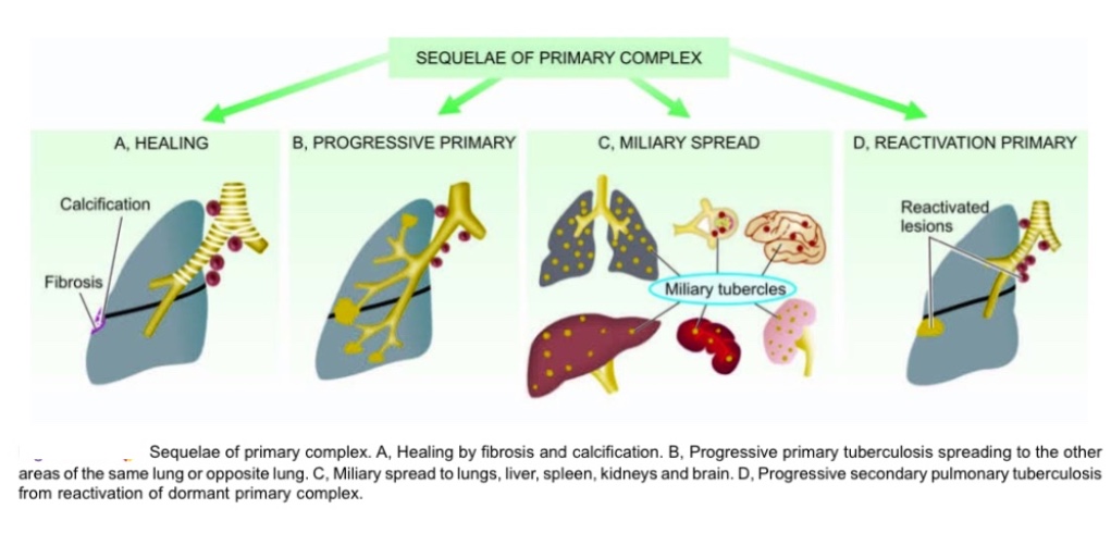
Autopsy of a 17 year old girl who died from pulmonary failure revealed a small area of caseous necrosis in the inferior lobe of the right lung, and occurrences of caseous necrosis in the bronchopulmonary, bronchial and bifurcational lymph nodes. What is the most probable postmortem diagnosis?
6. A 10 year old child had the mantoux tuberculin test administered. 48 hours later a papule up to 8 mm in diameter appeared on the site of the injection. What type of hypersensitivity reaction developed after the tuberculin injection?
7.
A 38 year old man died all of a sudden. Autopsy revealed myocardial infarction in the posterior wall of the left ventricle. What are the most likely alterations in myocardiocyte structure that can be revealed microscopically in the infarction focus?
8.
Histologic analysis of uterus mucous membrane revealed twisting glands, serrated and spinned, they were extended by stroma growth with proliferation of its cells. Formulate a diagnosis:
9.
Bacterioscopy of nasopharyngeal mucus taken from a 2,5 year old child with nasopharyngitis revealed gram- positive diplococci looking like coffee grains. What organs of the child are most likely to be affected if these microorganisms penetrate the blood?
10.
Autopsy of a man who died from ethylene glycol poisoning revealed that his kidneys are a little bit enlarged, edematic; their capsule can be easily removed. Cortical substance is broad and light-grey. Medullary substance is dark- red. What pathology had this man?
11.
Autopsy of a man who had tuberculosis revealed a 3x2 cm large cavity in the superior lobe of the right lung. The cavity was interconnected with a bronchus, its wall was dense and consisted of three layers: the internal layer was pyogenic, the middle layer was made by tuberculous granulation tissue and the external one was made by connective tissue. What is the most likely diagnosis?
12.
During examination of a 6-year-old child a doctor revealed greyish films on the pharyngeal tonsils. Their removal provoked moderate haemorrhage. Bacterioscopy revealed gram-positive clublike bacteria. What symptoms will develop in this child within the next few days if no specific treatment is provided?
13.
Autopsy of a woman with cerebral atherosclerosis revealed in the left cerebral hemisphere a certain focus that is presented by flabby, anhistic, greyish and yellowish tissue with indistinct edges. What pathological process is the case?
14.
A 38-year-old man died in the attempt of lifting weight. He had collaptoid state. Autopsy revealled an extensive aneurism rupture of thoracic aorta. He suffered from visceral syphilis during his lifetime. What pathological process caused weakness of aortic wall, its dilatation and rupture?
15.
Microscopical examination of an enlarged cervical lymph node revealed blurring of its structure, absence of lymphoid follicles; all the microscopic fields showed cells with roundish nuclei and thin limbus of basophil cytoplasm. It is known from the clinical data that other groups of lymph nodes are also enlarged as well as spleen and liver. What disease might be suspected?
16.
A worker of a cattle farm fell acutely ill and then died from the progressing intoxication. Autopsy revealed enlarged, hyposthenic spleen of dark-cherry colour when dissected; excessive pulp scraping. At the base and fornix of brain pia maters are edematous, soaked with blood, dark-red (\"scarlet hat\"). Microscopic examination revealed serous haemorrhagic inflammation of brain tissues and tunics along with destruction of small vessel walls. What is the most likely diagnosis?
Explanation
There are two medically important Bacillus species: Bacillus anthracis and Bacillus cereus. Bacillus anthracis causes anthrax.
Human disease occurs in 3 main forms: cutaneous, pulmonary (inhalation) and gastrointestinal. Humans are most often infected cutaneously at the time of trauma to the skin, which allows the spores on animal products such as hides, bristles and wool to enter. Spores can also be inhaled or when contaminated meat is ingested. After being inhaled, the organism moves rapidly to the mediastinal lymph nodes and causes hemorrhagic mediastinitis. Pathogenesis is based on the production of two exotoxins (Anthrax toxin) – edema factor and lethal factor. Hemorrhagic mediastinitis, septic shock hemorrhagic meningitis and death are severe life-threatening complications. In fatal cases, the organism may affect the spleen, liver, intestines, kidneys, adrenal glands and meninges.
Live spore vaccine (STI) is used for vaccination against anthrax.
STI live vaccine 17.
A man with a wound of his limb that had been suppurating for a long time died from intoxication. Autopsy revealed extreme emaciation, dehydration, brown atrophy of liver, myocardium, spleen and cross-striated muscles as well as renal amyloidosis. What diagnosis corresponds with the described presentations?
18. Chronic inflammation and transformation of the one-layer ciliated epithelium into multiple-layers flat epithelium was revealed in the thickened mucous membrane of the bronchus bioptate of the patient with smoke abuse. Which of the processes is the most likely?
19.
Microscopic examination of the enlarged neck gland of a 14 year old girl revealed destruction of the tissue structure of the node, absence of the lymph follicles, sclerotic areas and necrosis foci, cell constitution of the node is polymorphous, lymphocytes, eosinophils, big atypical cells with multilobular nuclei (Beresovsky-Sternberg cells) and mononuclear cells of the large size are present. What is the most likely diagnosis?
20. Pulmonary examination of a patient who has worked as a stone grinder for 9 years revealed small dense roundish nodules consisting of connective tissue. The nodules were found to have peripheral macrophages. Such pulmonary alterations are indicative of the following disease:
21. Autopsy of a 75-year-old man with a long history of atherosclerosis revealed a grey irregular-shaped focus of loose consistency in the right parietotemporal region of brain. What is the most likely cause of this process?
22.
Autopsy of a man with a malignant stomach tumour who had died from cancer intoxication revealed in the posteroinferior lung fields some dense, grayish-red irregular foci protruding above the section surface. Microscopic examination revealed exudate containing a large amount of neutrophils in the lumen and walls of small bronchi and alveoles. Such pulmonary alterations indicate the following disease:
23.
Autopsy of a 1,5-year-old child revealed haemorrhagic skin rash, moderate hyperaemia and edema of nasopharyngeal mucous membrane, small haemorrhages in the mucous membranes and internal organs; dramatic dystrophic alterations in liver and myocardium; acute necrotic nephrosis; massive haemorrhages in the adrenal glands. What disease are these alterations the most typical for?
Explanation
Morphologically, meningitis can be: meningococcal nasopharyngitis, meningococcal meningitis, meningococcemia. In meningococcemia, changes on the organs are characterized by generalized damage of microcirculation, skin rash, changes in the joints, vascular membrane of the eyes, adrenal glands and kidneys. Changes in the serous layers of the pericardium are observed. The rash is hemorrhagic, star-like, located mainly on the buttocks, lower extremities, eyelids and scleras. Focal necrosis and hemorrhages or bilateral massive hemorrhages with the development of acute adrenal insufficiency (waterhouse-friderichsen syndrome) are noted in the adrenals. Necrosis of nephrothelium of the tubules (necrotic nephrosis) is observed in the kidneys.
24.
A 46-year-old man had a bulging dark macula on skin that caused no discomfort. With time it began to increase in size and became painful. It turned dark brown and there was a nodule on palpation. Histological examination of tissues revealed spindle and polymorphous cells with multiple mitoses. Their cytoplasm contained brown pigment. What tumour is it?
25.
Autopsy of a 58 year old man revealed that bicuspid valve was deformed, thickened and unclosed. Microscopically: foci of collagen fibrilla are eosinophilic, react positively to fibrin. The most probably it is:
26.
Histological examination of the biopsy material obtained from the lower third of the esophagus of a 57-year-old male with the symptoms of continuous reflux revealed the change of the stratified squamous epithelium to the single-layer columnar glandular epithelium with signs of mucus production. Specify the pathological process in the mucous membrane:
27.
Microscopy of the bronchial wall revealed atrophy of the mucosa, metaplastic change from columnar to squamous epithelium, an increase in the number of goblet cells, diffuse infiltration of the bronchial wall with lymphoplasmacytic elements with a large number of neutrophilic granulocytes, pronounced sclerosis. Specify the morphological form of bronchitis:
28.
Microscopy of the myocardium of a patient who had died from heart failure revealed foci of fibrinoid necrosis located diffusely in the interstitial stroma, and often around the vessels. Such foci were surrounded by lymphocytes, macrophages, histiocytes. Pericardium was found to have signs of sero-fibrinous pericarditis. What is the most likely diagnosis?
29.
A 65-year-old male suddenly lost the vision in one eye due to the retinal detachment. The patient underwent enucleation. Histological examination of the removed eye retina and choroid revealed clusters of atypical cells with marked polymorphism of cells and nuclei, with a moderate number of mitoses including the pathological ones. The cell cytoplasm and intercellular medium contained brown pigment giving a positive DOPA reaction. Perls’ reaction was negative. What is the most likely diagnosis?
30.
At the post-mortem examination the stomach of a patient with renal failure was found to have a yellow-brown coating on the thickened mucosa.The coating was firmly adhering to its surface and had significant thickness. Microscopy revealed congestion and necrosis of mucosal and submucosal layers, fibrin presence. What is the most likely diagnosis?
31. Histological examination of the removed skin neoplasm revealed clusters and cords of atypical cells of stratified squamous epithelium, growing into the underlying tissue. What diagnosis can be assumed?
32.
Autopsy of a man who died from chronic cardiovascular collapse revealed \"tiger heart\". Sidewards of endocardium a yellowish-white banding can be seen; myocardium is dull, dark-yellow. What process caused this pathology?
33. Autopsy of a 62-year-old woman revealed a dense well-circumscribed node of 6 cm in diameter in the cranial cavity. The node was attached to the dura mater and histologically consisted of clusters and micro-concentric structures of endothelial cells, psammoma bodies. What kind of tumor was found at autopsy?
34.
Autopsy of a patient who suffered from croupous pneumonia and died from pneumococcal sepsis revealed 900 ml of turbid greenish-yellow liquid in the right pleural cavity. Pleural leaves are dull, plephoric. Name the clinicopathological form of inflammation in the pleural cavity:
35.
A patient died from progressive heart failure. Autopsy revealed that the heart was enlarged in diameter, flabby. The muscle section exhibited irregular blood supply. Histological study of myocardium revealed hyperemia, the stroma was found to have lymphohistiocytic infiltrates with degeneration of cardiomyocytes. The revealed morphological changes are indicative of:
36.
A patient underwent biopsy of the soft palate arches for a suspected tumor (macroscopy revealed an ulcer with a dense floor).Study of the biopsy material revealed mucosal necrosis with infiltration of lymphocytes, epithelioid cells, plasma cells, single neutrophils in the submucosa. There were also apparent signs of endovasculitis and perivasculitis. The described changes are typical for:
37. Examination of the removed stomach revealed a deep roundish defect with regular edges at the lesser curvature of the antrum. The defect reached the muscular tunic and was 1,5 cm in diameter. Within the defect floor there was a translucent dense area resembling of a hyaline cartilage. What process had developed in the floor of the stomach defect?
38.
A diseased child has a high fever, sore throat, swelling of submandibular lymph nodes. Objectively: pharyngeal mucosa is edematous, moderately hyperemic, the tonsils are enlarged, covered with grayish membrane tightly adhering to the tissues above. Attempts to remove the membrane produce the bleeding defects. What disease are these presentations typical for?
39.
14 days after quinsy a 15-year-old child presented with morning facial swelling, high blood pressure, \"meat slops\"urine. Immunohistological study of a renal biopsy sample revealed deposition of immune complexes on the basement membranes of the capillaries and in the glomerular mesangium. What disease developed in the patient?
40. Autopsy of a 78-year-old patient revealed that retroperitoneal tissue was soaked with blood, the abdominal aorta had a sacciform protrusion including a defect with irregular edges. The wall of the aorta was here and there of stone-like density. This is the complication of the following disease:
41.
Study of the biopsy material revealed a granuloma consisting of lymphocytes, plasma cells, macrophages with foamy cytoplasm (Mikulicz cells), many hyaline globules. What disease can you think of?
Explanation
Microscopic examination of specific granulomas:
* In Rhinoscleroma of nose, the granuloma (scleroma) consists of plasma cells, epitheloid cells, lymphocytes and hyaline sphere. Large macrophages with light cytoplasm containing klebsiella rhinoscleromatis (Mikulicz’s cells), sclerosis and hyalinosis takes place.
* In TB, the granuloma is reffered to as a tubercle and is classically characterized by the presence of central necrosis surrounded by epitheloid cells, lymphocytes, plasma cells and giant langhance cells. In contrast, caseous necrosis is rare in other granulomatous diseases.
* The syphilis granuloma is calle Gumma. Gumma consist of a central area of fibrinoid or caseous necrosis surrounded by mononuclear inflammatory cells, mostly plasma cells, lymphocytes, epitheloid cells and seldom-giant langhance cells. Around gumma forms the granulation tissue and endovasculitis.
* In Tuberculoid Leprosy, the epidermis contains confluent granulomas composed of macrophages, plasma cells and leprous Virchow’s cells – Leprous Virchow’s cells (or Leprous cells) refer to large foamy macrophages within fatty vacuoles containing leprous mycobacterium.
* Actinomycosis caused by Actinomyces. It occurs rarely in human but rather frequently in cattle. Characterized by the formation of painful abscess. Infected man often have poor oral hygiene or recent dental work. Does not form granulomas.
42. A child entering the school for the first time was given Mantoux test in order to determine if there was a need for revaccination. The reaction was negative. What is the meaning of this test result?
43.
At autopsy the occipital lobe of brain was found to have a cavity 2,5x1,5 cm large filled with a transparent liquid. The cavity had smooth brownish walls. What process had developed in the brain?
44.
A male patient is 28 years old. Histological study of a cervical lymph node revealed a change of its pattern due to the proliferation of epithelioid, lymphoid cells and macrophages having nuclei in form of a horseshoe. In the center of some cell clusters there were non-structured light-pink areas with fragments of nuclei. What disease are these changes typical for?
Explanation
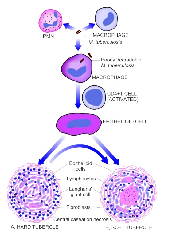
When tubercle bacilli are introduced into the tissue, they are ingested by the alveolar macrophage. Macrophages undergo changes resembling epithelial cells – EPITHELOID cells. Some of the macrophages form MULTINUCLEATED GIANT cells by fusion of adjacent cells (langerhan’s or foreign body type). The giant cells may have 20 or more nuclei. These nuclei may be arranged at the periphery like HORSE-SHOE, RING or clustered at the poles or they may be present centrally (foreign body giant cells). Lymphocytes, plasma cells and fibroblasts surround the epitheloid cells and giant cells (hard tubercle- no central necrosis). Within 10-14 days, the centre of the cellular mass begins to undergo caseation necrosis – soft tubercle. This is the hallmark of tuberculous lesions.
During the hematogenous spreading, the most serious immediate complication is MILIARY tuberculosis. The name miliary derives from its resemblance to millet seeds. Lesions of miliary tuberculosis consist of small granulomas, with a central necrotic portion. Organs often affected are lung, spleen, liver, kidney, meninges and bone marrow.
45. Bacteriological examination of purulent discharges from the urethra revealed some gram-negative bean- shaped bacteria located in the leukocytes. They can be identified as the causative agent of the following disease:
46. A 60-year-old patient with a long history of atherosclerosis and a previous myocardial infarction developed an attack of retrosternal pain. 3 days later the patient was hospitalized and then died of progressive cardiovascular insufficiency. At autopsy a white fibrous depressed area about 3 cm in diameter with clear boundaries was found in the posterior wall of the left ventricle and interventricular septum. The dissector evaluated these changes as:
47.
A 7-year-old boy got ill with diphtheria. On the third day he died of asphyxiation. At autopsy the mucosa of the larynx, trachea and bronchi had thickened, edematous, lustreless appearance and was covered with gray films which could be easily removed. Specify the type of laryngeal inflammation:
48.
A child with suspected tuberculosis was given Mantoux test. After 24 hours the site of the allergen injection got swollen, hyperemic and painful. What are the main components that determine such response of the body?
Explanation
Mantoux test is a type IV Hypersensitivity reaction (HSR), which involves macrophages,T-lymphocytes and lymphokines (cytokines). Mononuclear cells (lymphocytes, monocytes, macrophages).
Remember,it is antibody independent (i.e does not involve antibodies).
B-lymphocytes - Plasma cells - Ig(Antibodies)------- none is involved in Type IV HSR.
The Mantoux skin test should be read between 48 and 72hrs after administration. The basis of reading is the presence or absence of induration, which may be determined by inspection and by palpation. A record should also be made of formation of vesicles, bullae, lymphangitis, ulceration and necrosis at the test site. The formation of vesicles, bullae or necrosis at the test site indicates positive result. A negative mantoux result usually signifies that the individual has never been exposed to Mycobacterium tuberculosis i.e. absence of cell mediated immunity to tuberculin.
49. Histological examination of biopsy samples taken from the thickened edges of a gastric ulcer revealed small clusters of small, markedly atypical hyperchromatic epithelial cells that were localized in the overdeveloped stroma. Specify the tumor:
50.
A 10-year-old child was found to have a congenital hypoplasia of the left kidney. Ultrasound examination revealed that the right kidney was markedly enlarged and had regular shape. No functional disorders were revealed. Specify the process that developed in the right kidney:
51.
A 35-year-old female patient has undergone biopsy of the breast nodules. Histological examination has revealed enhanced proliferation of the small duct and acini epithelial cells, accompanied by the formation of glandular structures of various shapes and sizes, which were located in the fibrous stroma. What is the most likely diagnosis?
52.
A 54-year-old female was brought to the casualty department after a car accident. A traumatologist diagnosed her with multiple fractures of the lower extremities. What kind of embolism is most likely to develop in this case?
53.
There are cortical and medullary substances separated by connective tissue layer in the endocrine gland specimen. Parenchyma cells make up three zones in cortical substance, with rounded masses in the superficial zone, parallel chords in the middle one, reticular structure of cell chords in the deep one. What gland is it?
54. A patient underwent surgical removal of a cavitary liver lesion 2 cm in diameter. It was revealed that the cavity wall was formed by dense fibrous connective tissue; the cavity contained muddy, thick, yellowish-greenish fluid with an unpleasant odor. Microscopically, the fluid consisted mainly of polymorphonuclear leukocytes. What pathological process are these morphological changes typical for?
55.
Autopsy has revealed shrunken kidneys weighing 50 mg, with fine-grained surface and uniformly thinned substance. Microscopic investigation has shown the thickening of arteriole walls due to accumulation of homogeneous anhistic pink-coloured masses in them. Glomerules were undersized, sclerotic, with atrophied tubules. What disease are these changes characteristic of?
56.
On the 24th day since the onset of disease, a male patient diagnosed with typhoid fever and undergoing treatment in an infectious diseases hospital has suddenly developed clinical presentations of acute abdomen leading to the death of the patient. During autopsy peritonitis has been revealed, with numerous ulcers covering the colon mucosa and reaching as deep as muscular and, in places, serous tunic. The ulcers have smooth edges and even floor. The intestinal wall is perforated. What stage of typhoid fever has the lethal complication arisen at?
57.
An HIV-positive patient’s cause of death is acute pulmonary insufficiency resulting from pneumonia. Pathohistological investigation of lungs has revealed interstitial pneumonia, alveolocyte desquamation and their methamorphoses: alveolocyte enlargement, large intranuclear inclusions surrounded by lightly-colored areas. Transformed cells resemble owl’s eye. Name the pneumonia causative agent:
58.
A 37-year-old male patient developed pseudoarthrosis after a closed fracture of the femur. Specify the type of regeneration in the patient:
59.
A patient with suspected tumor of lung had been admitted to the oncological department. Examination revealed localised pathology in the inferior lobe of the left lung. How many bronchopulmonary segments does this lobe have?
60. Tissue sampling of a 37-year-old
male patient with chronic renal disease has revealed the following: sclerosis, lymphocytic and plasmocytic infiltration of renal pelvis and calices walls, dystrophy and atrophy of tubules. Remaining tubules are enlarged and stretched with colloid masses, epithelium is flattened out (\"scutiform\" or \"shield-shaped\" kidney). What is the most likely diagnosis?
61.
Autopsy of a 50-year-old male who had tuberculosis revealed a dense gray-white nidus in form of a nodule 2 cm in diameter in the subpleural portion of the upper right lobe. The pleura in this region was thickened, in the pleural cavity there was a small amount of serous hemorrhagic fluid. Histological study of the region revealed some glandular structures with signs of cellular atypia and abnormal mitoses, which were found within the fibrous connective tissue. What other pathology had developed in the lungs?
62.
A 10-year-old child has painful swallowing, neck edema, temperature rise up to 39, 0oC, the whole body is covered with bright-red petechial rash. Back of the throat and tonsils are hyperemic, the tongue is crimson-colored. Tonsillar surface is covered with isolated grayish-colored necrosis nidi. What disease is it?
63. A 3-year-old child with meningeal symptoms died. Postmortem macroscopy of the pia matter revealed miliary nodules which were microscopically represented by a focus of caseous necrosis with masses of epithelioid and lymphoid cells with some crescent-shaped large cells inbetween having peripheral nuclei. Specify the type of meningitis in the child:
Explanation
When tubercle bacilli are introduced into the tissue, they are ingested by the alveolar macrophage. Macrophages undergo changes resembling epithelial cells – EPITHELOID cells. Some of the macrophages form MULTINUCLEATED GIANT cells by fusion of adjacent cells (langerhan’s or foreign body type). The giant cells may have 20 or more nuclei. These nuclei may be arranged at the periphery like HORSE-SHOE, RING or clustered at the poles or they may be present centrally (foreign body giant cells). Lymphocytes, plasma cells and fibroblasts surround the epitheloid cells and giant cells (hard tubercle- no central necrosis). Within 10-14 days, the centre of the cellular mass begins to undergo caseation necrosis – soft tubercle. This is the hallmark of tuberculous lesions.
During the hematogenous spreading, the most serious immediate complication is MILIARY tuberculosis. The name miliary derives from its resemblance to millet seeds. Lesions of miliary tuberculosis consist of small granulomas, with a central necrotic portion. Organs often affected are lung, spleen, liver, kidney, meninges and bone marrow.
64. X-ray examination of a patient allowed to diagnose a tumor in the superior lobe of the right lung. There is a probability of metastases spread to the following lymph nodes:
65.
A young woman suddenly developed fever up to 39oC accompanied by a strong headache. Examination revealed marked nuchal rigidity. Spinal puncture was performed. Gram-stained smear of cerebrospinal fluid contained many neutrophils and Gram-positive diplococci. What bacteria could be the cause of this disease?
66. A patient has hoarseness of voice. During laryngoscopy a gray-white larynx tumor with papillary surface has been detected. Microscopic investigation has shown the following: growth of connective tissue covered with multilayer, strongly keratinized pavement epithelium, no cellular atypia. What is the most likely diagnosis?
67.
During autopsy approximately 2,0 liters of pus have been found in the abdominal cavity of the corpse. Peritoneum is lustreless and has grayish shade, serous tunic of intestines has grayish-colored coating that is easily removable. Specify the most likely type of peritonitis in the patient:
68.
Autopsy of the dead patient who died from pulmonary edema revealed a large yellow-grey nidus in the myocardium, and a fresh thrombus in the coronary artery. What is the most likely diagnosis?
69. A male patient is 28 years old. Histological study of a cervical lymph node revealed a change of its pattern due to the proliferation of epithelioid, lymphoid cells and macrophages having nuclei in form of a horseshoe. In the center of some cell clusters there were non-structured light-pink areas with fragments of nuclei. What disease are these changes typical for?
70.
During autopsy the following has been revealed: the meninges of the upper cerebral hemispheres are extremely plethoric, of yellow-green color and are soaked with purulent effluent. What kind of meningitis is characterised by such clinical presentations?
71. A 40-year-old patient with the progressing staphylococcal purulent periodontitis developed purulent inflammation of bone marrow spaces of the alveolar process, and then of the body of mandible. Microscopy revealed thinning of bone trabeculae, foci of necrosis, bone sequesters surrounded by the connective tissue capsule. What is the most likely diagnosis?
72.
A 40-year-old female patient has undergone thyroidectomy. Histological study of thyroid gland found the follicles to be of different size and contain foamy colloid, follicle epithelium is high and forms papillae, there is focal lymphocytic infiltration in stroma. Diagnose the thyroid gland disease:
73.
Extensive thromboembolic infarction of the left cerebral hemispheres, large septic spleen, immunocomplex glomerulonephritis, ulcers on the edges of the aortic valves, covered with polypous thrombus with colonies of staphylococcus were revealed on autopsy of the young man who died in coma. What disease caused cerebral thromboemboly?
74.
A 45 year old male died from disseminated tuberculosis. On autopsy the symptoms of tuberculosis were confirmed by both microscopical and histological analyses. All the affected organs had epithelioid cell granulomas with caseous necrosis in the centre. What kind of hypersensitivity reaction underlies the process of granuloma development?
Explanation
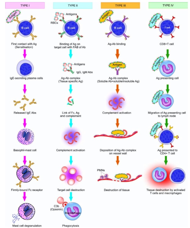
Type I (Immediate, Anaphylaxis, Reagin): IgE (immunoglobulin E)-dependent activation of mast cells/basophils, usually accompanied by eosinophilia e.g. urticaria (hives), hay fever, asthma (wheezing), rhinitis and conjunctivitis (stuffy nose and itchy eyes; usually seasonal)
Type II (cytotoxic): antibody dependent reactions e.g. Goodpasture syndrome, Myasthenia gravis, Graves disease, ABO hemolytic disease of newborn etc.
Type III (immune-complex): deposition of antigen-antibody complexes e.g. systemic lupus erythromatous (SLE), Arthus reaction, serum sickness, poststreptococcal glomerulonephritis etc.
Type IV (cell mediated, delayed): antibody-independent T-cell mediated reactions e.g. positive mantoux reaction (tuberculin test), hashimoto’s thyroiditis or transplant rejection etc.
75.
A patient with high-titer antinuclear antibodies died from progressing renal impairment. Autopsy revealed mesangioproliferative glomerulonephritis and abacterial polypous endocarditis. There was periarterial bulbar sclerosis in spleen and productive proliferative vasculitis in skin. What is the most likely diagnosis?
76.
A 50 year old patient underwent resection of tumour of large intestine wall. Microscopically it presents itself as fascicles of divergent collagen fibers of different thickness and form and some monomorphous fusiform cells that are irregularly distributed among the fibers. Cellular atypia is not evident. What tumour is it?
77. A patient who abuses smoking has chronic bronchitis. Biopsy of his primary bronchus revealed multilayer pavement epithelium. What pathological process was revealed in the bronchus?
78. 2 days after labour a woman developed shock along with DIC syndrome that caused her death. Autopsy revealed purulent endomyometritis, regional purulent lymphangitis, lymphadenitis and purulent thrombophlebitis. There were also dystrophic alterations and interstitial inflammation of parenchymal organs. What is the most likely diagnosis?
79.
Acute renal impairment caused death of a bleeding patient. Autopsy revealed enlarged kidneys with a broad pale pink cortical layer expressively demarcated from dark red renal pyramids. Macroscopic examination revealed lack of epithelial nuclei of convoluted tubules, tubulorrhexis, phlebostasis. The cell nuclei of choroid glomus and straight tubules were present. What pathology is it?
80.
Examination of a bronchial tissue sample revealed atrophy of mucous membrane, cystic degeneration of glands, focal metaplastic changes of lining prismatic epithelial cells into multilayer squamous cells; increase in goblet cell number; in some parts of bronchial wall and especially in the mucous membrane there was marked cellular inflammatory infiltration and growth of granulation tissue bulging into the bronchial lumen in form of a polyp. What is the most likely diagnosis?
81.
Autopsy of a 75 year old patient who had been suffering from disseminated atherosclerosis and died under chronic cardiac failure revealed constriction and deformation of coronary arteries, tuberous intima whose section appeared to be white and petrosal. Specify the stage of atherosclerosis morphogenesis:
82. A 71 year old man had been presenting with diarrhea for 10 days. The feces had admixtures of blood and mucus. He was delivered to a hospital in grave condition and died 2 days later. Autopsy of the body revealed the following: diphtheritic colitis with multiple irregularly-shaped ulcers of different depth in both sigmoid colon and rectus. Bacteriological analysis revealed Shigella. What was the main disease?
83.
A 46-year-old man complains of difficulties with nasal breathing. Mikulicz’s cells, accumulation of epithelioid cells, plasmocytes, lymphocytes, hyaline balls were discovered in the biopsy material of the thickened nasal mucosa. What is the most likely diagnosis?
Explanation
Microscopic examination of specific granulomas:
* In Rhinoscleroma of nose, the granuloma (scleroma) consists of plasma cells, epitheloid cells, lymphocytes and hyaline sphere. Large macrophages with light cytoplasm containing klebsiella rhinoscleromatis (Mikulicz’s cells), sclerosis and hyalinosis takes place.
* In TB, the granuloma is reffered to as a tubercle and is classically characterized by the presence of central necrosis surrounded by epitheloid cells, lymphocytes, plasma cells and giant langhance cells. In contrast, caseous necrosis is rare in other granulomatous diseases.
* The syphilis granuloma is calle Gumma. Gumma consist of a central area of fibrinoid or caseous necrosis surrounded by mononuclear inflammatory cells, mostly plasma cells, lymphocytes, epitheloid cells and seldom-giant langhance cells. Around gumma forms the granulation tissue and endovasculitis.
* In Tuberculoid Leprosy, the epidermis contains confluent granulomas composed of macrophages, plasma cells and leprous Virchow’s cells – Leprous Virchow’s cells (or Leprous cells) refer to large foamy macrophages within fatty vacuoles containing leprous mycobacterium.
84.
A young man has a painless formation without marked borders in the soft tissues of his thigh. On the tissue bioptate the formation looks like flesh of fish and consists of immature fibroblast-like cells with multiple mitosis growing through the muscles. What is the most likely diagnosis?
85.
Autopsy of a man ill with severe hypothyroidism revealed that connective tissue, organ stroma, adipose and cartilaginous tissues were swollen, semi- transparent, mucus-like. Microscopic examination of tissues revealed stellate cells having processes with mucus between them. What type of dystrophy is it?
86. A 28 year old patient had high arterial pressure, hematuria and facial edemata. In spite of treatment renal insufficiency was progressing. 6 months later the patient died from uremia. Microscopic examination of his kidneys and their glomerules revealed proliferation of capsule nephrothelium and of podocytes with \"demilune\" formation, sclerosis and hyalinosis of glomerules. What disease corresponds with the described picture?
87.
A 6 year old child was delivered to a hospital. Examination revealed that the child couldn’t fix his eyes, didn’t keep his eyes on toys, eye ground had the cherry-red spot sign. Laboratory analyses showed that brain, liver and spleen had high rate of ganglioside glycometide. What congenital disease is the child ill with?
88. Autopsy of a 49-year-old woman who died from chronic renal insufficiency, revealed: kidneys were dense, reduced, multicoloured, with haemorrhagic areas. Microscopic examination revealed some hematoxylin bodies in the nuclei of the renal tubule epithelium; \"wire-loop\" thickening of the glomerular capillary basement membrane; here and there in the capillaries some hyaline thrombi and foci of fibrinoid necrosis were present. What is the most likely diagnosis?
89.
A 20 year old patient died from intoxication 8 days after artificial illegal abortion performed in her 14-15th week of pregnancy. Autopsy of the corpse revealed yellowish colour of eye sclera and of skin, necrotic suppurative endometritis, multiple pulmonary abscesses, spleen hyperplasia with a big number of neutrophils in its sinuses. What complication after abortion was developed?
90. Histological examination of a skin tissue sampling revealed granulomas consisting of macrophagal nodules with lymphocytes and plasmatic cells. There are also some big macrophages with fatty vacuoles containing causative agents of a disease packed up in form of spheres (Virchow\'s cells). Granulation tissue is well vascularized. What disease is this granuloma typical for?
91.
Examination of a young woman revealed a tumour up to 3 cm in diameter in form of a knot localized along the acoustic nerve. The tumour is homogenous, soft and elastic, of pink-and-white colour. Microscopically the tumour contains clusters of cells with oval nuclei. Fibrous cell clusters form regular structures made up by parallel rows of regularly oriented cells arranged in form of a palisade. Zones between the rows of cells are acellular and homogenous (Verocai bodies). What tumour is it?
92.
A section of the left lung was found to have an area of dense red tissue. The area was cone-shaped, stood out distinctly from the healthy tissue, with its base directed to the pleura. The dissected tissue was granular, dark-red. What is the most likely diagnosis?
93. A patient with severe course of respiratory viral infection presented with clinical signs of progressing heart failure that led to his death in the 2nd week of disease. Autopsy revealed that the heart cavities were significantly dilated, the heart was flabby. Histological examination of the myocardium revealed microvascular plethora and diffuse stroma infiltration with lymphocytes and histiocytes. What is the most likely diagnosis?
94. A pathology-histology laboratory received a vermiform appendix up to 2,0 cm thick. Its serous membrane was pale, thick and covered with yellowish-green films. The wall was flaccid, of grayish-red colour. The appendix lumen was dilated and filled with yellowish-green substance. Histological examination revealed that the appendix wall was infiltrated with neutrophils. Specify the appendix disease:
95. Gynecological examination of the uterine cervix in a 30-year-old woman revealed some bright-red lustrous spots that easily bleed when touched. Biopsy showed that a part of the uterine cervix was covered with cylindrical epithelium with papillary outgrowths; in the depth of tissue the growth of glands was present. What pathology of the uterine cervix was revealed?
96. Autopsy of a man who died from influenza revealed that the heart was slightly enlarged and pastose. The surface of the incision of myocardium appeared to be pale, with specks. Microscopic examination revealed signs of parenchymatous adipose and hydropic degeneration, edematic stroma with scant lymphocytic and macrophage infiltration; plethoric vessels; perivascular petechial hemorrhages. What type of myocarditis is it?
97. Autopsy of a man, who had been suffering from the multiple bronchiectasis for 5 years and died from chronic renal insufficiency, revealed that kidneys were dense and enlarged, with thickened cortical layer of white colour with greasy lustre. What renal disease might be suspected?
98.
Autopsy of a 50-year-old man revealed the following changes: his right lung was moderately compact in all parts, the dissected tissue was found to be airless, fine-grained, dryish. Visceral pleura had greyish-brown layers of fibrin. What is the most likely diagnosis?
99. A man had worked in a coal mine for over 20 years. After his death autopsy revealed that his lungs were dense, grayish-black and had large areas of neogenic connective tissue containing a lot of microphages with black pigment in the cytoplasm. What is the most likely diagnosis?
100.
Colonoscopy of a patient with dysentery revealed that the mucous membrane of the large intestine was hyperemic, edematic, and its surface was covered with grey-and-green layerings. What morphological form of dysenteric colitis is it?
101.
An 8-year-old child was admitted to the infectious department with fever (up to 38oC ) and punctuate bright-red skin rash. The child was diagnosed as having scarlet fever. Objectively: mucous membrane of pharynx is apparently hyperaemic and edematic, the tonsils are enlarged and have dull yellowish-grey foci with some black areas. What inflammation is the reason for the pharynx alterations?
102.
While examining a patient an otolaryngologist noticed hyperaemia and significantly edematous tonsils with a grayish film upon them. Microscopical examination of this film revealed some gram-positive bacilli placed at an angle with each other. What disease might be suspected?
103.
Examination of a patient revealed a dense, movable skin tumour that is standing out distinctly from the surrounding tissues. Its section is found to be white and composed of fibrous tissue. Microscopic examination revealed interlacing collagen fibers and few cells. What tumour is it?
104. A 2 year old child had acute respiratory viral infection and died from cardiopulmonary decompensation. Autopsy revealed that his right lung was hyperemic; in the 2nd, 6th and 10th segments and on the incision there were airless yellowish foci of irregular form, from several mm up to 1 cm large. Microscopical examination revealed exudate consisting mainly of neutrophils in the given areas of pulmonary tissue in the alveoles, bronchioles and bronchial tubes. What is the most probable diagnosis?
105. A 23 year old man has perforation of hard palate. In the area of this perforation there was a compact well-defined formation. Microscopic examination of the resected formation revealed a large focus of caseous necrosis surrounded by granulation tissue with endovasculitis, cellular infiltration composed of lymphocytes, epithelioid cells (mainly plasmocytes). What is the most probable diagnosis?
106.
A patient who has been abusing tobacco smoking for a long time has got cough accompanied by excretion of viscous mucus; weakness after minor physical stress, pale skin. The patient has also lost 12,0 kg of body weight. Endoscopic examination of biopsy material his illness was diagnosed as squamous cell carcinoma. Name a pathological process that preceded formation of the tumour:
107.
A 33 year old man died from uraemia. Autopsy revealed enlarged kidneys weighing 500,0 each and consisting of multiple cavities 0,5-2 cm in diameter. The cavities were full of light-yellow transparent liquid. Renal pelvis and ureters had no peculiarities. What renal disease caused uraemia?
108.
A patient died from acute cardiac insufficiency, among clinical presentations there was gastrointestinal haemorrhage. Examination of mucous membrane of stomach revealed some defects reaching myenteron; their edges and bottom were mostly even and loose, some of them contained dark-red blood. What pathological process was revealed?
109.
Examination of a 66 year old patient revealed a lytic tumour in the locus of pathological rib fracture. Histologically this tumour consists of atypical plasmoblasts. Further examination revealed osteoporosis in the bones of vertebral column and pelvis. These changes are typical for:
110. A patient had been suffering from profuse diarrhea and vomiting for 2 days. He died from acute dehydration. Autopsy revealed that the intestinal wall was edematic and hyperemic, with multiple haemorrhages in the mucous membrane. Intestine lumen contains whitish fluid resembling of rice water. What disease caused death?
111.
Autopsy of a 5 year old child revealed in the area of vermis of cerebellum a soft greyish-pink node 2 cm in diameter with areas of haemorrhage. Histologically this tumour consisted of atypical monomorphous small roundish cells with big polymorphous nuclei. What tumour is it?
112. A 46 year old patient who had been suffering from tuberculosis for 6 years died from massive pulmonary haemorrhage. Autopsy revealed different-sixed foci of sclerosis and caseous necrosis in lungs, in the upper part of the right lung there was a cavity 5 cm in diameter with dense grey walls, the cavity contained liquid blood and blood clots. What type of tuberculosis is it?
113. A patient ill with thrombophlebitis of his lower limbs had chest pain, blood spitting, progressing respiratory insufficiency that led to his death. Autopsy diagnosed multiple lung infarctions. What is the most probable cause of their development?ow
114.
A 4 year old child complained of pain during deglutition, indisposition. Objectively: palatine arches and tonsils are moderately edematic and hyperemic, there are greyish-white films up to 1 mm thick closely adhering to the subjacent tissues. What pathological process are these changes typical for?
115.
A patient has been suffering from diarrhea for 5 day. On the fifth day colonoscopy revealed that membrane of rectum was inflamed, there were greyish-green films closely adhering to the subjacent tissue. What is the most probable diagnosis?
116.
48 hours after tuberculine test (Mantoux test) a child had a papule 10 mm in diameter on the spot of tuberculine injection. What hypersensitivity mechanism underlies these changes?
117.
6 months after labour a woman had uterine hemorrhage. Gynaecological examination of uterine cavity revealed a dark-red tissue with multiple cavities resembling of a \"sponge\". Microscopic examination of a tumour revealed in blood lacunas atypic light epithelial Langhans cells and giant cells of syncytiotrophoblast. What tumour is it?
118.
A 30 year old man had been suffering from acute respiratory disease and died from cardiopulmonary decompensation. Autopsy revealed fibrinous-haemorrhagic inflammation in the mucous membrane of larynx and trachea, destructive panbronchitis, enlarged lungs that look black due to the multiple abcesses, haemorrhages, necrosis. What is the most probable postmortem diagnosis?
119.
Autopsy of a man who died from burn disease revealed brain edema, liver enlargement as well as enlargement of kidneys with wide light-grey cortical layer and plethoric medullary area. Microscopic examination revealed necrosis of tubules of main segments along with destruction of basal membranes, intersticium edema with leukocytic infiltration and haemorrhages. What is the most probable postmortem diagnosis?
120.
Examination of coronary arteries revealed atherosclerotic calcified plaques closing vessel lumen by 1/3. The muscle has multiple whitish layers of connective tissue. What process was revealed in the myocardium?
121.
A 22 year old patient from the West Ukraine complains of laboured nasal breathing. Morphological examination of biopsy material of nasal mucous membrane revealed lymphoid, epitheliod, plasma cells as well as Mikulicz’s cells. What is the most probable diagnosis?
Explanation
Microscopic examination of specific granulomas:
* In Rhinoscleroma of nose, the granuloma (scleroma) consists of plasma cells, epitheloid cells, lymphocytes and hyaline sphere. Large macrophages with light cytoplasm containing klebsiella rhinoscleromatis (Mikulicz’s cells), sclerosis and hyalinosis takes place.
* In TB, the granuloma is reffered to as a tubercle and is classically characterized by the presence of central necrosis surrounded by epitheloid cells, lymphocytes, plasma cells and giant langhance cells. In contrast, caseous necrosis is rare in other granulomatous diseases.
* The syphilis granuloma is calle Gumma. Gumma consist of a central area of fibrinoid or caseous necrosis surrounded by mononuclear inflammatory cells, mostly plasma cells, lymphocytes, epitheloid cells and seldom-giant langhance cells. Around gumma forms the granulation tissue and endovasculitis.
* In Tuberculoid Leprosy, the epidermis contains confluent granulomas composed of macrophages, plasma cells and leprous Virchow’s cells – Leprous Virchow’s cells (or Leprous cells) refer to large foamy macrophages within fatty vacuoles containing leprous mycobacterium.
122. Examination of the anterior abdominal wall of a pregnant woman revealed a tumour-like formation that arose on the spot of a tumour that was removed two years ago. The neoplasm was well-defined, dense, 2х1 cm large. Histological examination revealed that the tumour was composed of differentiated connective tissue with prevailing collagen fibres. What tumour might be suspected?
123.
The liver puncture biopsy of a patient with hepatocellular insufficiency revealed hydropic and ballooning degenerationof hepatocytes, necrosis of certain cells, presence of Kaunsilmen’s bodies. Portal and lobular stroma were infiltrated mostly with lymphocytes and macrophages as well as with a small number of polymorphonuclear lymphocytes. What is the most likely diagnosis?
124.
A patient with tuberculosis died from progressing cardiopulmonary decompensation. Autopsy in the region of the right lung apex revealed a cavity 5 cm in diameter communicating with lumen of a segmental bronchus. On the inside cavity walls are covered with caseous masses with epithelioid and Langhans cells beneath them. What morphological form of tuberculosis is it?
125.
A 70-year-old male patient died from acute coronary insufficiency. He had knee joint swelling, gonycampsis and gonalgia during his lifetime. Pathomorphologic examination of the deformed joints and synovial membranes revealed membrane hyperemia with multiple perivascular inflammatory infiltrations made by lymphocytes, plasmocytes and macrophagocytes. There was an accumulation of organized fibrin covering some areas of synovium membrane and looking like rice grains in the articular liquid. What is the most likely diagnosis?
126. A 49-year-old patient with croupous pneumonia died from pneumococcal septicemia. Autopsy revealed up to 700 ml of turbid greenish-yellow foul-smelling liquid in the left pleural cavity. The pleural leaflets were dull and plethoric. What form of pleural inflammation is it?
127.
A patient died from cardiopulmonary decompensation. Histological examination revealed diffused pulmonary affection along with interstitial edema, infiltration of tissue by limphocytes, macrophages, plasmocytes; pulmonary fibrosis, panacinar emphysema. What is the most likely diagnosis?
128.
Acute renal impairment caused death of a bleeding patient. Autopsy revealed enlarged kidneys with a broad pale pink cortical layer expressively demarcated from dark red renal pyramids. Macroscopic examination revealed lack of epithelial nuclei of convoluted tubules, tubulorrhexis, phlebostasis. The cell nuclei of choroid glomus and straight tubules were present. What pathology is it?
129.
Medical examination of a 20-year-old woman revealed a dense incapsulated node 1 cm in diameter that was palpated in the mammary gland. The postoperative biopsy revealed connective tissue overgrowth around the mammary ducts and glandular components of different diameter that didn’t make lobules and bore no signs of cellular abnormality. What diagnosis will be made?